
Deposition Date
2003-09-03
Release Date
2004-03-23
Last Version Date
2024-10-30
Entry Detail
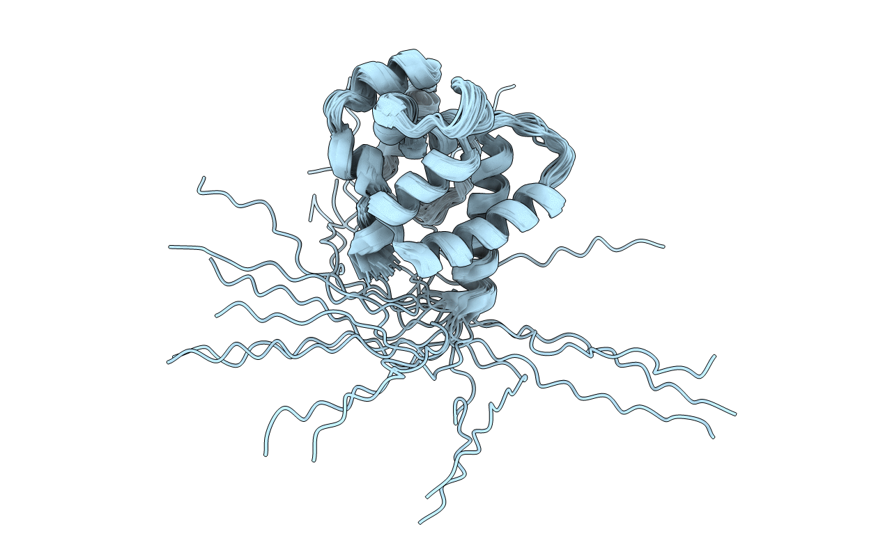
PDB ID:
1QWV
Keywords:
Title:
Solution structure of Antheraea polyphemus pheromone binding protein (ApolPBP)
Biological Source:
Source Organism(s):
Antheraea polyphemus (Taxon ID: 7120)
Expression System(s):
Method Details:
Experimental Method:
Conformers Calculated:
200
Conformers Submitted:
20
Selection Criteria:
structures with the lowest energy
