1QWG image
Deposition Date 2003-09-02
Release Date 2003-12-09
Last Version Date 2024-02-14
Entry Detail
PDB ID:
1QWG
Keywords:
Title:
Crystal structure of Methanococcus jannaschii phosphosulfolactate synthase
Biological Source:
Source Organism:
Host Organism:
Method Details:
Experimental Method:
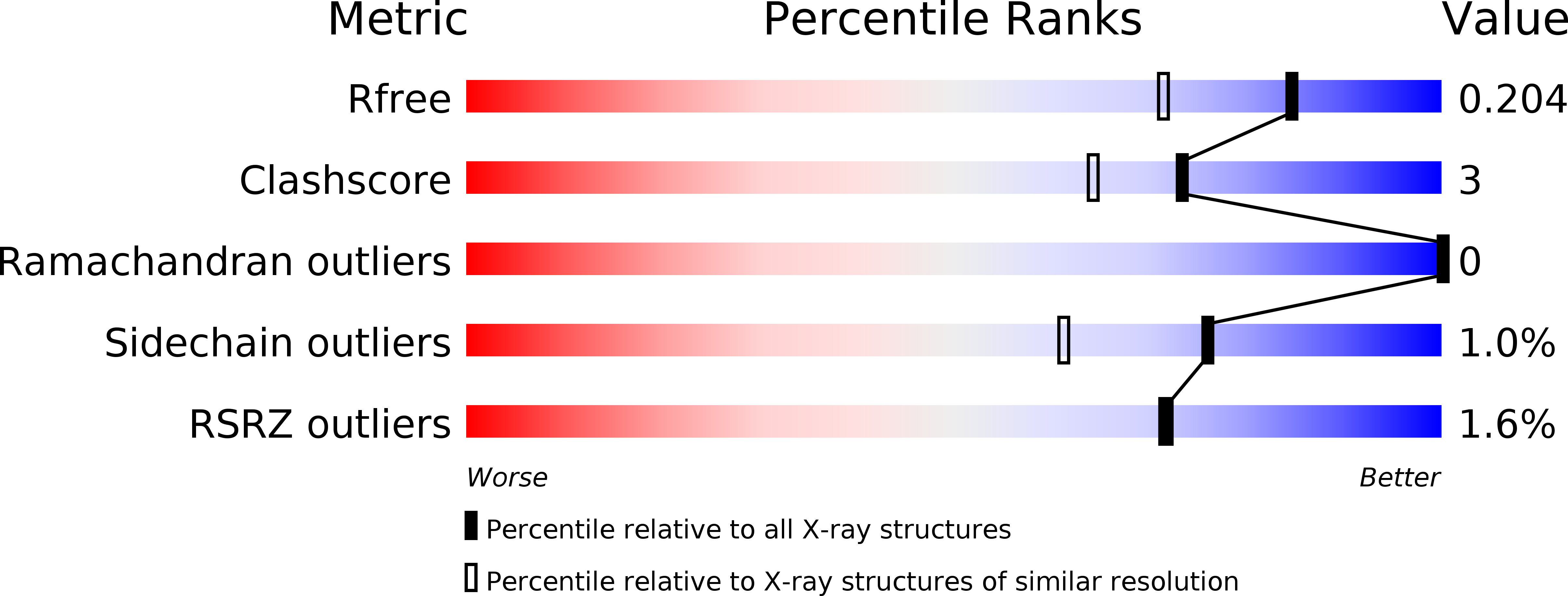
Resolution:
1.60 Å
R-Value Free:
0.19
R-Value Work:
0.17
R-Value Observed:
0.17
Space Group:
P 21 3
Macromolecular Entities
Polymer Type:polypeptide(L)
Molecule:(2R)-phospho-3-sulfolactate synthase
Gene (Uniprot):comA
Chain IDs:A
Chain Length:251
Number of Molecules:1
Biological Source:Methanocaldococcus jannaschii
Ligand Molecules
Primary Citation
The structural determination of phosphosulfolactate synthase from Methanococcus jannaschii at 1.7-A resolution: an enolase that is not an enolase
J.Biol.Chem. 278 45858 45863 (2003)
PMID: 12952952 DOI: 10.1074/jbc.M307486200

Abstact

Members of the enolase mechanistically diverse superfamily catalyze a wide variety of chemical reactions that are related by a common mechanistic feature, the abstraction of a proton adjacent to a carboxylate group. Recent investigations into the function and mechanism of the phosphosulfolactate synthase encoded by the ComA gene in Methanococcus jannaschii have suggested that ComA, which catalyzes the stereospecific Michael addition of sulfite to phosphoenolpyruvate to form phosphosulfolactate, may be a member of the enolase superfamily. The ComA-catalyzed reaction, the first step in the coenzyme M biosynthetic pathway, likely proceeds via a Mg2+ ion-stabilized enolate intermediate in a manner similar to that observed for members of the enolase superfamily. ComA, however, has no significant sequence similarity to any known enolase. Here we report the x-ray crystal structure of ComA to 1.7-A resolution. The overall fold for ComA is an (alpha/beta)8 barrel that assembles with two other ComA molecules to form a trimer in which three active sites are created at the subunit interfaces. From the positions of two ordered sulfate ions in the active site, a model for the binding of phosphoenolpyruvate and sulfite is proposed. Despite its mechanistic similarity to the enolase superfamily, the overall structure and active site architecture of ComA are unlike any member of the enolase superfamily, which suggests that ComA is not a member of the enolase superfamily but instead acquired an enolase-type mechanism through convergent evolution.

Legend

Protein

Chemical

Disease

Primary Citation of related structures