Deposition Date
1999-06-30
Release Date
2000-07-05
Last Version Date
2024-11-13
Entry Detail
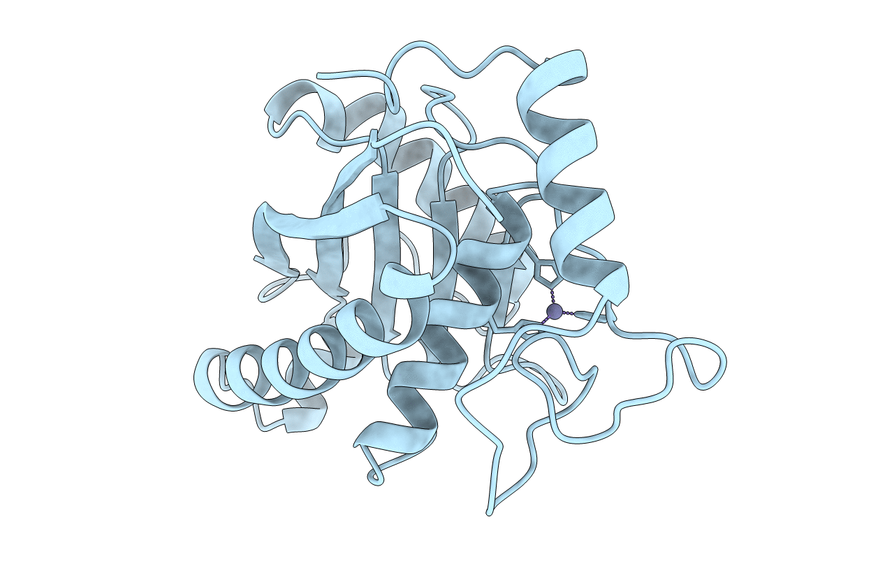
PDB ID:
1QUA
Keywords:
Title:
CRYSTAL STRUCTURE OF ACUTOLYSIN-C, A HEMORRHAGIC TOXIN FROM THE SNAKE VENOM OF AGKISTRODON ACUTUS, AT 2.2 A RESOLUTION
Biological Source:
Source Organism(s):
Deinagkistrodon acutus (Taxon ID: 36307)
Method Details:
Experimental Method:
Resolution:
2.20 Å
R-Value Free:
0.27
R-Value Work:
0.17
R-Value Observed:
0.17
Space Group:
P 21 21 21