
Deposition Date
1999-06-25
Release Date
1999-07-07
Last Version Date
2024-11-06
Entry Detail
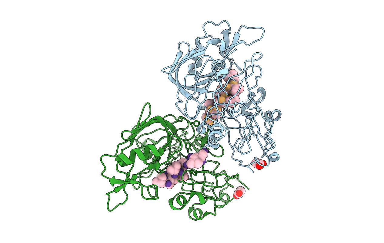
PDB ID:
1QS8
Keywords:
Title:
Crystal structure of the P. vivax aspartic proteinase plasmepsin complexed with the inhibitor pepstatin A
Biological Source:
Source Organism(s):
Plasmodium vivax (Taxon ID: 5855)
Streptomyces argenteolus subsp. toyonakensis (Taxon ID: 285516)
Streptomyces argenteolus subsp. toyonakensis (Taxon ID: 285516)
Expression System(s):
Method Details:
Experimental Method:
Resolution:
2.50 Å
R-Value Free:
0.25
R-Value Work:
0.19
R-Value Observed:
0.19
Space Group:
P 41 21 2
