
Deposition Date
1999-06-16
Release Date
1999-07-06
Last Version Date
2024-05-22
Entry Detail
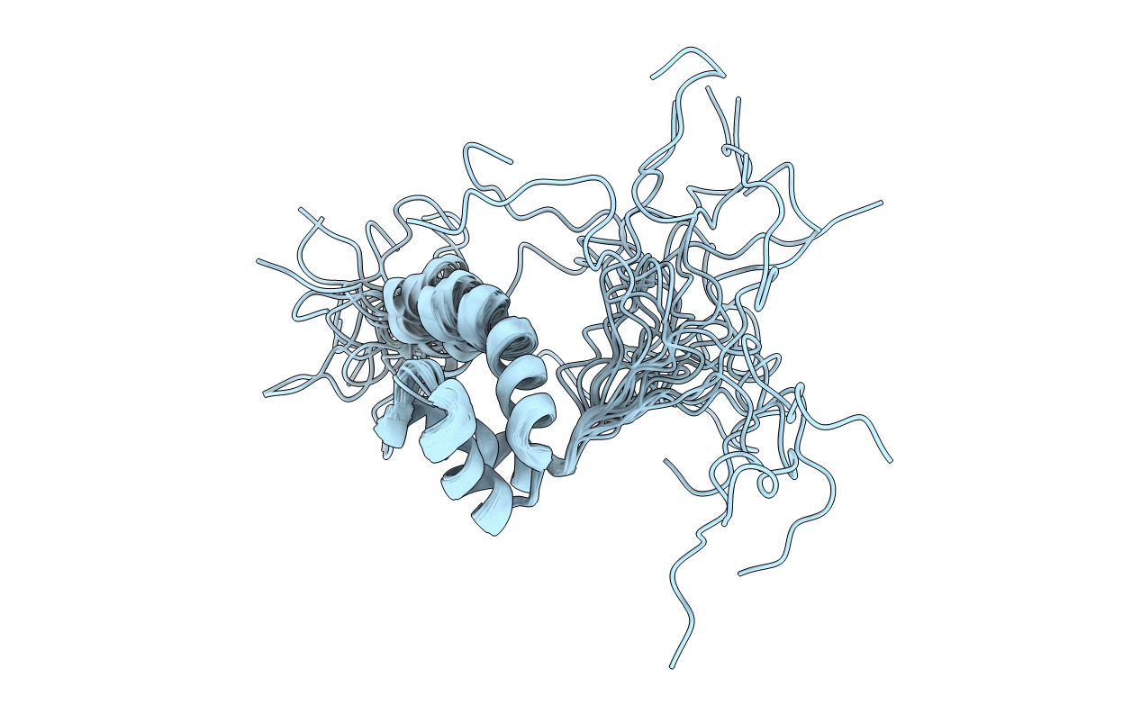
PDB ID:
1QRY
Keywords:
Title:
Homeobox protein VND (ventral nervous system defective protein)
Biological Source:
Source Organism(s):
Drosophila melanogaster (Taxon ID: 7227)
Expression System(s):
Method Details:
Experimental Method:
Conformers Calculated:
100
Conformers Submitted:
20
Selection Criteria:
STRUCTURES WITH ACCEPTABLE COVALENT GEOMETRY,STRUCTURES WITH THE LEAST RESTRAINT VIOLATIONS,STRUCTURES WITH THE LOWEST ENERGY
