1QQQ image
Deposition Date 1999-06-07
Release Date 1999-06-14
Last Version Date 2024-10-30
Entry Detail
PDB ID:
1QQQ
Keywords:
Title:
CRYSTAL STRUCTURE ANALYSIS OF SER254 MUTANT OF ESCHERICHIA COLI THYMIDYLATE SYNTHASE
Biological Source:
Source Organism:
Escherichia coli (Taxon ID: 562)
Host Organism:
Method Details:
Experimental Method:
Resolution:
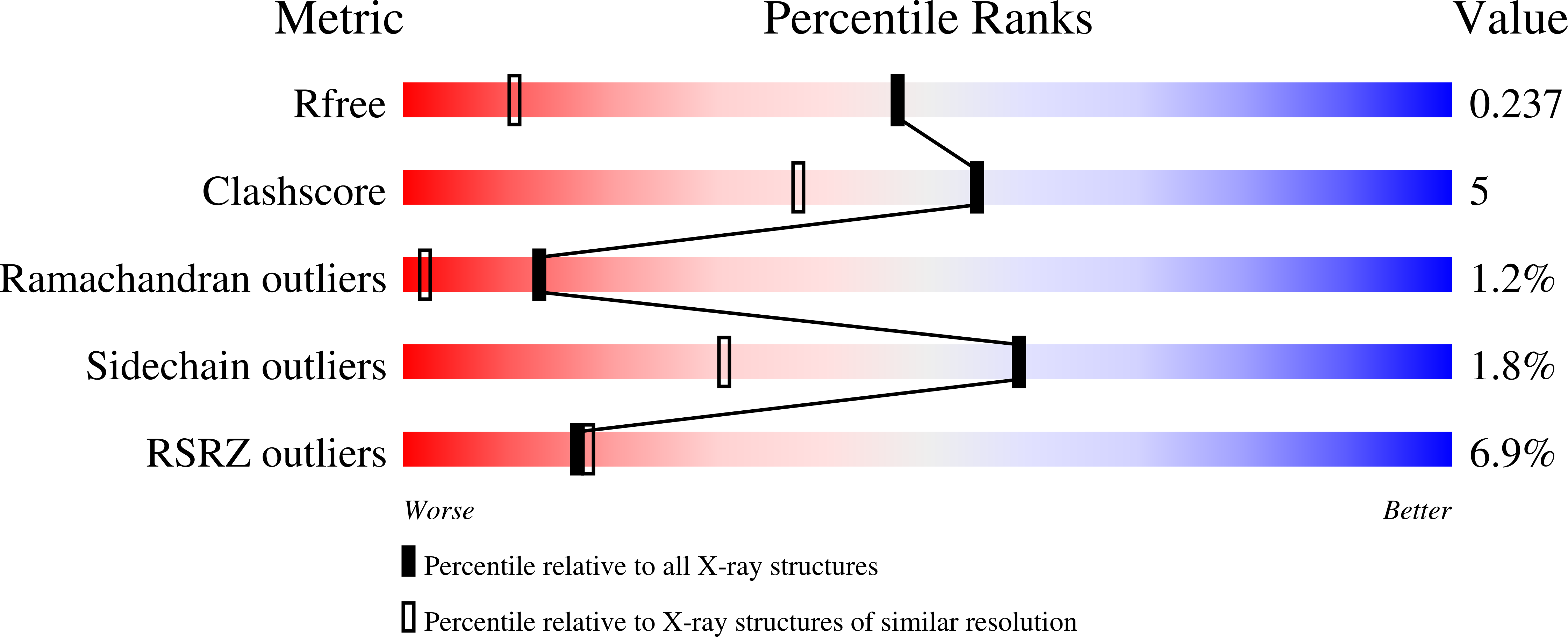
1.50 Å
R-Value Free:
0.23
R-Value Work:
0.22
R-Value Observed:
0.22
Space Group:
I 21 3
Macromolecular Entities
Polymer Type:polypeptide(L)
Molecule:THYMIDYLATE SYNTHASE
Gene (Uniprot):thyA
Mutations:P254S
Chain IDs:A
Chain Length:264
Number of Molecules:1
Biological Source:Escherichia coli
Modified Residue
Compound ID Chain ID Parent Comp ID Details 2D Image
CME A CYS S,S-(2-HYDROXYETHYL)THIOCYSTEINE
CXM A MET N-CARBOXYMETHIONINE
Ligand Molecules
Primary Citation
Drug-resistant variants of Escherichia coli thymidylate synthase: effects of substitutions at Pro-254.
Mol.Pharmacol. 57 359 366 (2000)
PMID: 10648646

Abstact

Drug-resistant variants of thymidylate synthase (TS) can potentially be used in gene therapy applications to decrease the myelosuppressive side effects of TS-directed anticancer agents or to select genetically modified cells in vivo. Mutations of proline 303 of human TS confer resistance to TS-directed fluoropyrimidines and antifolates (). We generated the corresponding variants in Escherichia coli TS (ecTS), position 254, to better understand the mechanism by which mutations at this residue confer resistance. In addition, because ecTS is intrinsically resistant to several antifolates when compared with human TS, we suspected that greater resistance could be achieved with the bacterial enzyme. The P254L enzyme conferred >100-fold resistance to both raltitrexed and 5-fluoro-2'-deoxyuridine (FdUrd) compared with wild-type ecTS. Four additional mutants (P254F, P254S, P254G, and P254D), each of which complemented growth of a TS-deficient cell line, were generated, isolated, and characterized. Steady-state values of K(m) for dUMP and k(cat) were not substantially different among the variants and were comparable with the wild-type values, but K(m) for methylenetetrahydrofolate (CH(2)H(4)PteGlu) was >10-fold higher for P254D. Values of k(on) and k(off) for nucleotide binding, which were obtained by stopped-flow spectroscopy, were virtually unchanged among the mutants. Drastic differences were observed for CH(2)H(4)PteGlu binding, with K(d) values >15-fold higher than observed with the wild-type enzyme; surprisingly, the proposed isomerization reaction that is very evident for the wild-type enzyme is not observed with P254S. The decrease in affinity for CH(2)H(4)PteGlu correlates well with K(i) values obtained for three TS-directed inhibitors. These results show that mutations at Pro-254 specifically affect the initial binding interactions between enzyme and cofactor and also alter the ability of the mutant enzymes to undergo conformational changes that occur on ternary complex formation. The crystal structure of P254S was determined at 1.5 A resolution and is the most precise structure of TS available. When compared with wild-type TS, the structure shows local conformational changes affecting mostly Asp-253; its carbonyl is rotated approximately 40 degrees, and the side chain forms an ion pair with Arg-225.

Legend

Protein

Chemical

Disease

Primary Citation of related structures