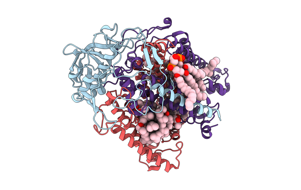
1QOV image
Entry Detail
PDB ID:
1QOV
Title:
PHOTOSYNTHETIC REACTION CENTER MUTANT WITH ALA M260 REPLACED WITH TRP (CHAIN M, A260W)
Biological Source:
Source Organism:
PDB Version:
Deposition Date:
1999-11-17
Release Date:
1999-12-13
Method Details:
Experimental Method:
Resolution:
2.10 Å
R-Value Free:
0.18
R-Value Work:
0.16
Space Group:
P 31 2 1
Macromolecular Entities
Polymer Type:polypeptide(L)
Description:PHOTOSYNTHETIC REACTION CENTER
Mutations:YES
Chain IDs:A (auth: H)
Chain Length:260
Number of Molecules:1
Biological Source:RHODOBACTER SPHAEROIDES
Polymer Type:polypeptide(L)
Description:PHOTOSYNTHETIC REACTION CENTER
Mutations:YES
Chain IDs:B (auth: L)
Chain Length:281
Number of Molecules:1
Biological Source:RHODOBACTER SPHAEROIDES
Polymer Type:polypeptide(L)
Description:PHOTOSYNTHETIC REACTION CENTER
Mutations:YES
Chain IDs:C (auth: M)
Chain Length:307
Number of Molecules:1
Biological Source:RHODOBACTER SPHAEROIDES
Primary Citation
Structural Details of an Interaction between Cardiolipin and an Integral Membrane Protein
Proc.Natl.Acad.Sci.USA 96 14706 ? (1999)
PMID: 10611277 DOI: 10.1073/PNAS.96.26.14706

Abstact

Anionic lipids play a variety of key roles in biomembrane function, including providing the immediate environment for the integral membrane proteins that catalyze photosynthetic and respiratory energy transduction. Little is known about the molecular basis of these lipid-protein interactions. In this study, x-ray crystallography has been used to examine the structural details of an interaction between cardiolipin and the photoreaction center, a key light-driven electron transfer protein complex found in the cytoplasmic membrane of photosynthetic bacteria. X-ray diffraction data collected over the resolution range 30.0-2.1 A show that binding of the lipid to the protein involves a combination of ionic interactions between the protein and the lipid headgroup and van der Waals interactions between the lipid tails and the electroneutral intramembrane surface of the protein. In the headgroup region, ionic interactions involve polar groups of a number of residues, the protein backbone, and bound water molecules. The lipid tails sit along largely hydrophobic grooves in the irregular surface of the protein. In addition to providing new information on the immediate lipid environment of a key integral membrane protein, this study provides the first, to our knowledge, high-resolution x-ray crystal structure for cardiolipin. The possible significance of this interaction between an integral membrane protein and cardiolipin is considered.

Legend

Protein

Chemical

Disease

Primary Citation of related structures