1QOQ image
Deposition Date 1999-11-15
Release Date 2000-11-10
Last Version Date 2023-12-13
Entry Detail
PDB ID:
1QOQ
Keywords:
Title:
CRYSTAL STRUCTURE OF WILD-TYPE TRYPTOPHAN SYNTHASE COMPLEXED WITH INDOLE GLYCEROL PHOSPHATE
Biological Source:
Source Organism:
Host Organism:
Method Details:
Experimental Method:
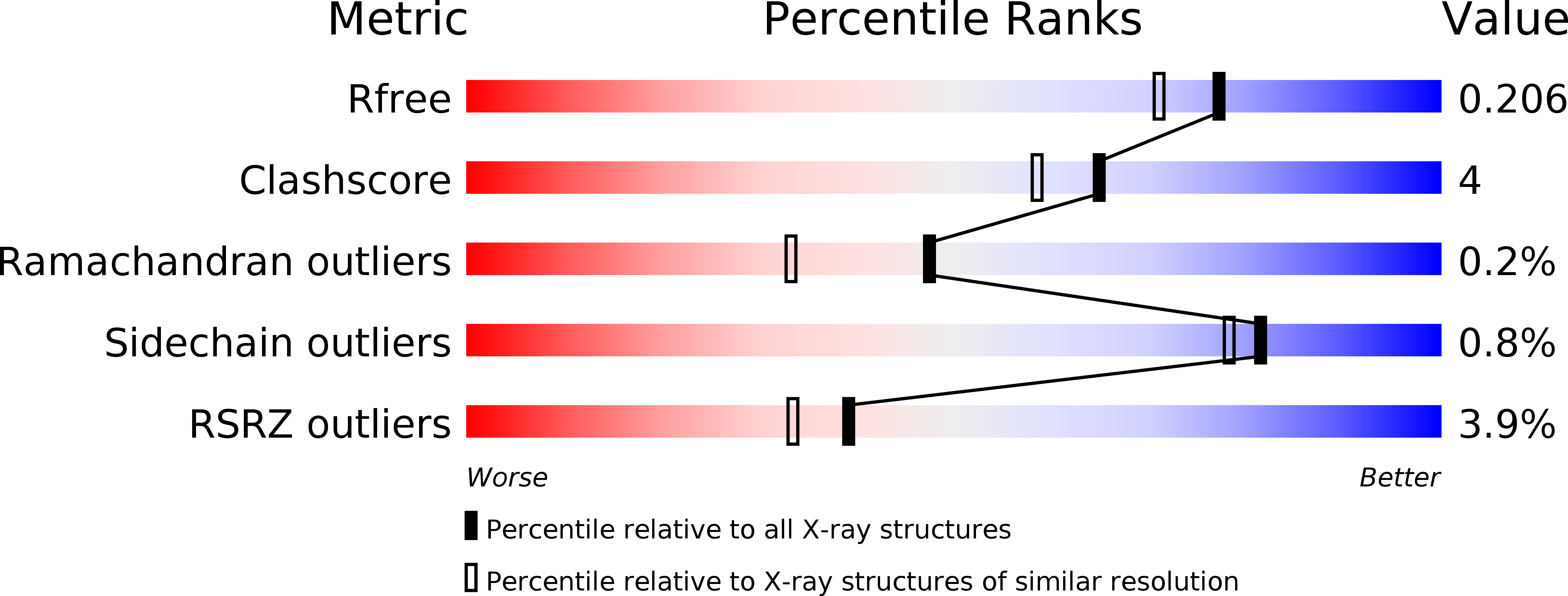
Resolution:
1.80 Å
R-Value Free:
0.21
R-Value Work:
0.17
Space Group:
C 1 2 1
Macromolecular Entities
Polymer Type:polypeptide(L)
Molecule:TRYPTOPHAN SYNTHASE ALPHA CHAIN
Gene (Uniprot):trpA
Chain IDs:A
Chain Length:268
Number of Molecules:1
Biological Source:SALMONELLA TYPHIMURIUM
Polymer Type:polypeptide(L)
Molecule:TRYPTOPHAN SYNTHASE BETA CHAIN
Gene (Uniprot):trpB
Chain IDs:B
Chain Length:396
Number of Molecules:1
Biological Source:SALMONELLA TYPHIMURIUM
Primary Citation
Crystal Structure of Wild-Type Tryptophan Synthase Complexed with the Natural Substrate Indole-3-Glycerol Phosphate.
Biochemistry 38 16469 ? (1999)
PMID: 10600108 DOI: 10.1021/BI9920533

Abstact

We used freeze trapping to stabilize the Michaelis complex of wild-type tryptophan synthase and the alpha-subunit substrate indole-3-glycerol phosphate (IGP) and determined its structure to 1. 8 A resolution. In addition, we determined the 1.4 A resolution structure of the complex with indole-3-propanole phosphate (IPP), a noncleavable IGP analogue. The interaction of the 3'-hydroxyl of IGP with the catalytic alphaGlu49 leads to a twisting of the propane chain and to a repositioning of the indole ring compared to IPP. Concomitantly, the catalytic alphaAsp60 rotates resulting in a translocation of the COMM domain [betaGly102-betaGly189, for definition see Schneider et al. (1998) Biochemistry 37, 5394-5406] in a direction opposite to the one in the IPP complex. This results in loss of the allosteric sodium ion bound at the beta-subunit and an opening of the beta-active site, thereby making the cofactor pyridoxal 5'-phosphate (PLP) accessible to solvent and thus serine binding. These findings form the structural basis for the information transfer from the alpha- to the beta-subunit and may explain the affinity increase of the beta-active site for serine upon IGP binding.

Legend

Protein

Chemical

Disease

Primary Citation of related structures