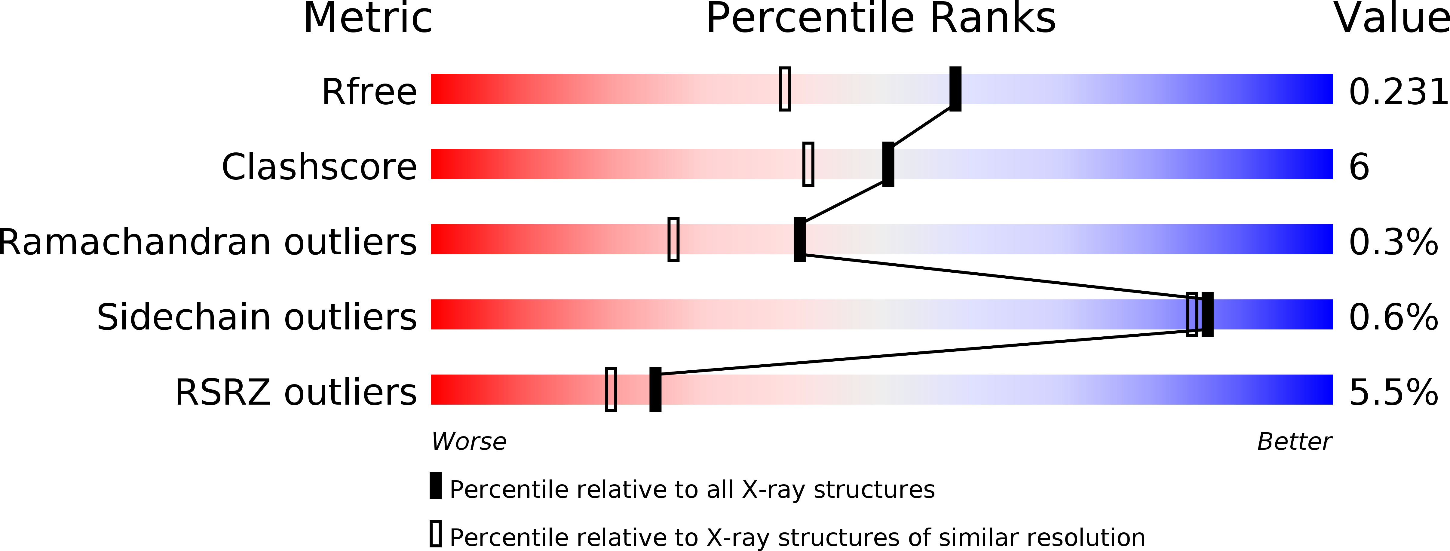
1QO7 image
Deposition Date 1999-11-04
Release Date 2000-02-10
Last Version Date 2024-05-08
Entry Detail
PDB ID:
1QO7
Keywords:
Title:
Structure of Aspergillus niger epoxide hydrolase
Biological Source:
Source Organism:
Host Organism:
Method Details:
Experimental Method:
Resolution:
1.80 Å
R-Value Free:
0.23
R-Value Work:
0.22
R-Value Observed:
0.22
Space Group:
P 1 21 1
Macromolecular Entities
Structures with similar UniProt ID
Protein Blast
Polymer Type:polypeptide(L)
Molecule:EPOXIDE HYDROLASE
Gene (Uniprot):hyl1
Mutagens:YES
Chain IDs:A, B
Chain Length:394
Number of Molecules:2
Biological Source:ASPERGILLUS NIGER
Primary Citation
Structure of Aspergillus Niger Epoxide Hydrolase at 1.8A Resolution: Implications for the Structure and Function of the Mammalian Microsomal Class of Epoxide Hydrolases
Structure 8 111 ? (2000)
PMID: 10673439 DOI: 10.1016/S0969-2126(00)00087-3

Abstact

BACKGROUND: Epoxide hydrolases have important roles in the defense of cells against potentially harmful epoxides. Conversion of epoxides into less toxic and more easily excreted diols is a universally successful strategy. A number of microorganisms employ the same chemistry to process epoxides for use as carbon sources. RESULTS: The X-ray structure of the epoxide hydrolase from Aspergillus niger was determined at 3.5 A resolution using the multiwavelength anomalous dispersion (MAD) method, and then refined at 1.8 A resolution. There is a dimer consisting of two 44 kDa subunits in the asymmetric unit. Each subunit consists of an alpha/beta hydrolase fold, and a primarily helical lid over the active site. The dimer interface includes lid-lid interactions as well as contributions from an N-terminal meander. The active site contains a classical catalytic triad, and two tyrosines and a glutamic acid residue that are likely to assist in catalysis. CONCLUSIONS: The Aspergillus enzyme provides the first structure of an epoxide hydrolase with strong relationships to the most important enzyme of human epoxide metabolism, the microsomal epoxide hydrolase. Differences in active-site residues, especially in components that assist in epoxide ring opening and hydrolysis of the enzyme-substrate intermediate, might explain why the fungal enzyme attains the greater speeds necessary for an effective metabolic enzyme. The N-terminal domain that is characteristic of microsomal epoxide hydrolases corresponds to a meander that is critical for dimer formation in the Aspergillus enzyme.

Legend

Protein

Chemical

Disease

Primary Citation of related structures