1QNP image
Deposition Date 1999-10-20
Release Date 2000-10-19
Last Version Date 2024-11-06
Entry Detail
PDB ID:
1QNP
Keywords:
Title:
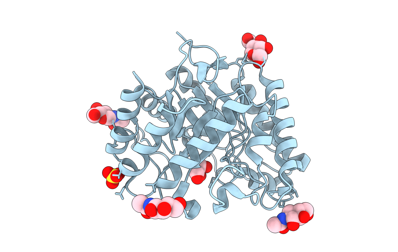
The 3-D structure of a Trichoderma reesei b-mannanase from glycoside hydrolase family 5
Biological Source:
Source Organism:
Host Organism:
Method Details:
Experimental Method:
Resolution:
1.50 Å
R-Value Free:
0.17
R-Value Work:
0.12
Space Group:
P 1 21 1
Macromolecular Entities
Polymer Type:polypeptide(L)
Molecule:ENDO-1,4-B-D-MANNANASE
Gene (Uniprot):man1
Chain IDs:A
Chain Length:344
Number of Molecules:1
Biological Source:TRICHODERMA REESEI
Modified Residue
Compound ID Chain ID Parent Comp ID Details 2D Image
ASN A ASN GLYCOSYLATION SITE
Primary Citation
The Three-Dimensional Structure of a Trichoderma Reesei Beta-Mannanase from Glycoside Hydrolase Family 5.
Acta Crystallogr.,Sect.D 56 3 ? (2000)
PMID: 10666621 DOI: 10.1107/S0907444999013943

Abstact

The crystal structure of the catalytic core domain of beta-mannanase from the fungus Trichoderma reesei has been determined at a resolution of 1.5 A. The structure was solved using the anomalous scattering from a single non-isomorphous platinum complex with two heavy-metal sites in space group P2(1). The map computed with the experimental phases was enhanced by the application of an automated model building and refinement procedure using the amplitudes and experimental phases as observations. This approach is expected to be of more general application. The structure of the native enzyme and complexes with Tris-HCl and mannobiose are also reported: the mannobiose binds in subsites +1 and +2. The structure is briefly compared with that of the homologous beta-mannanase from the bacterium Thermomonospora fusca.

Legend

Protein

Chemical

Disease

Primary Citation of related structures