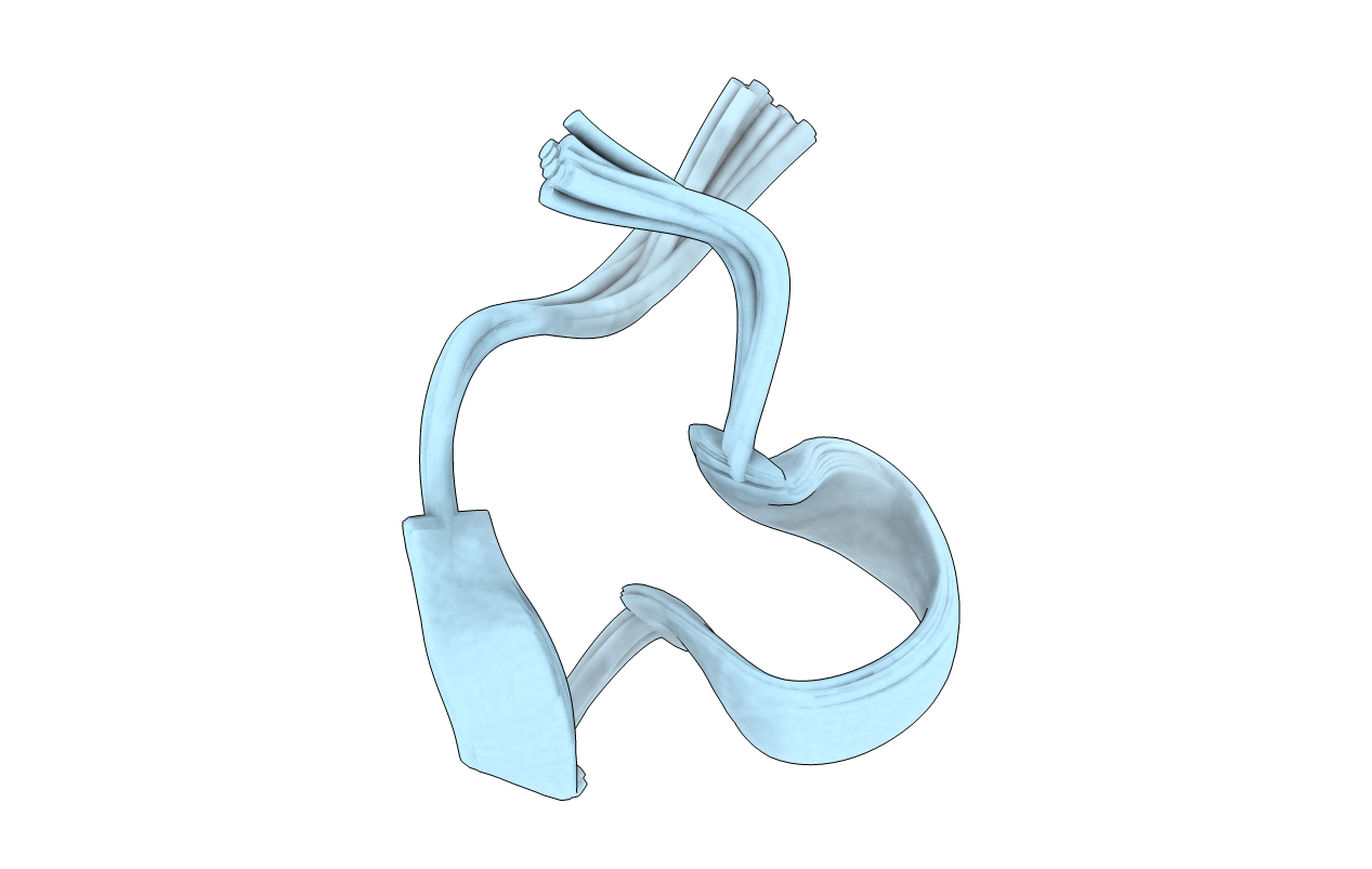
1QMW image
Entry Detail
PDB ID:
1QMW
Keywords:
Title:
Solution structure of alpha-conotoxin SI
Biological Source:
Source Organism:
PDB Version:
Deposition Date:
1999-10-08
Release Date:
2000-08-25
Method Details:
Experimental Method:
Conformers Calculated:
50
Conformers Submitted:
36
Selection Criteria:
LOWEST ENERGY STRUCTURES
Macromolecular Entities
Polymer Type:polypeptide(L)
Description:ALPHA-CONOTOXIN SI
Chain IDs:A
Chain Length:14
Number of Molecules:1
Biological Source:CONUS STRIATUS
Ligand Molecules
Primary Citation
Solution Structure of Alpha-Conotoxin Si
FEBS Lett. 476 287 ? (2000)
PMID: 10913630 DOI: 10.1016/S0014-5793(00)01724-5

Abstact

The nuclear magnetic resonance solution structure of alpha-conotoxin SI has been determined at pH 4.2. The 36 lowest energy structures show that alpha-conotoxin SI exists in a single major solution conformation and is stabilized by six hydrogen bonds. Comparisons are made between the SI solution structure and the solution and crystal structures of alpha-conotoxin GI. Surprisingly, a high degree of similarity between the backbone conformations of the GI crystal and the SI solution structures is seen in the region of lowest sequence homology, namely residues Gly-8 to Ser-12. This similarity is more surprising when considering that in SI a proline replaces the Arg-9 found in GI. The correspondence in conformation in this region provides the definitive evidence that it is the loss of the arginine basic charge at residue 9 which determines the differences in toxicity between GI and SI, rather than any changes in conformation induced by the cyclic proline residue.

Legend

Protein

Chemical

Disease

Primary Citation of related structures