
Deposition Date
1999-09-21
Release Date
1999-09-27
Last Version Date
2023-12-13
Entry Detail
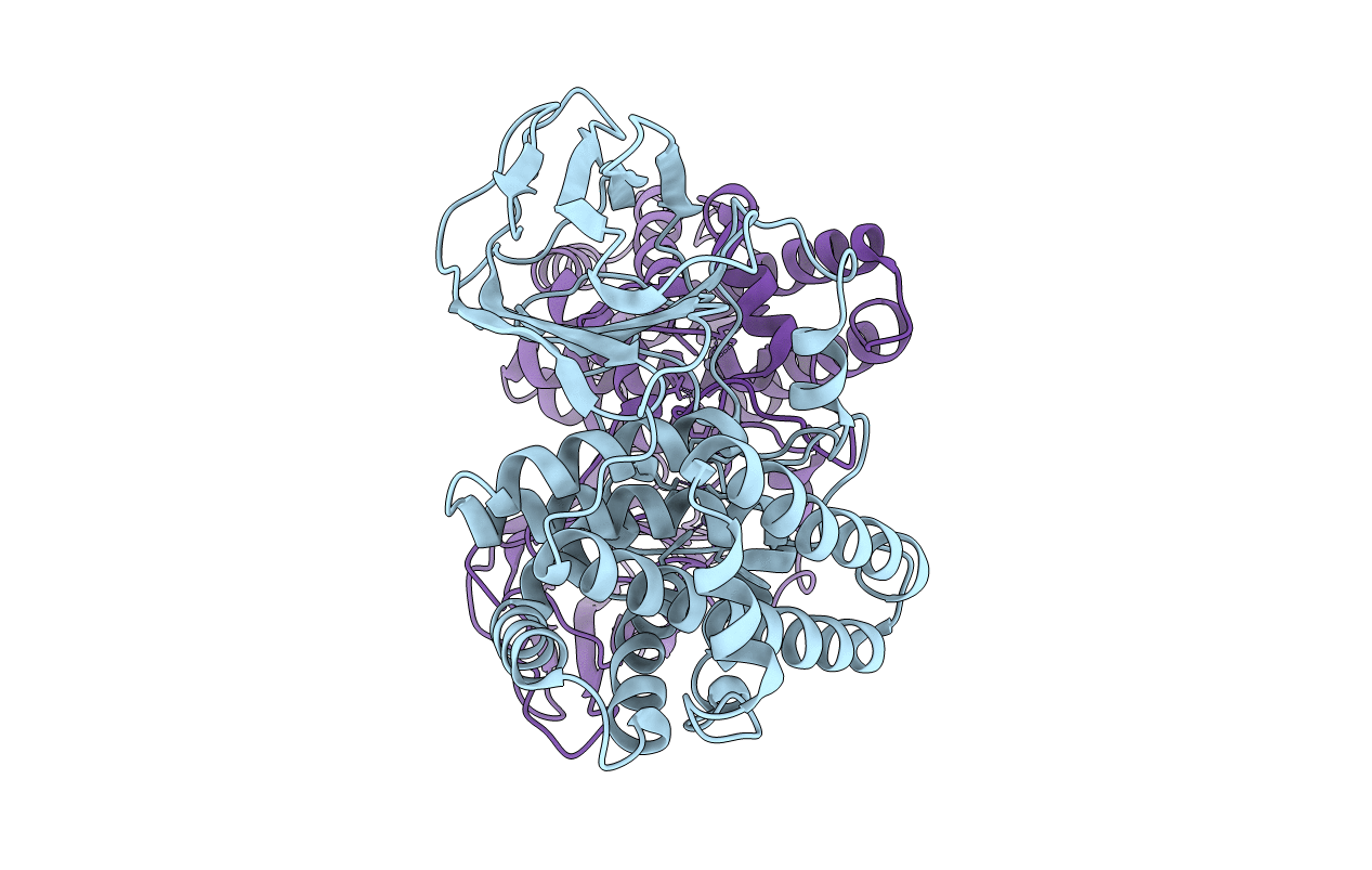
PDB ID:
1QM6
Keywords:
Title:
Closed form of Clostridium perfringens alpha-toxin strain NCTC8237
Biological Source:
Source Organism(s):
CLOSTRIDIUM PERFRINGENS (Taxon ID: 1502)
Expression System(s):
Method Details:
Experimental Method:
Resolution:
2.50 Å
R-Value Free:
0.24
R-Value Work:
0.22
R-Value Observed:
0.22
Space Group:
H 3 2
