
Deposition Date
1999-09-11
Release Date
2000-09-14
Last Version Date
2024-10-16
Entry Detail
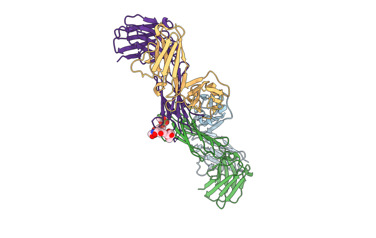
PDB ID:
1QLR
Keywords:
Title:
CRYSTAL STRUCTURE OF THE FAB FRAGMENT OF A HUMAN MONOCLONAL IgM COLD AGGLUTININ
Biological Source:
Source Organism(s):
HOMO SAPIENS (Taxon ID: 9606)
Method Details:
Experimental Method:
Resolution:
2.83 Å
R-Value Free:
0.26
R-Value Work:
0.21
Space Group:
P 31 2 1
