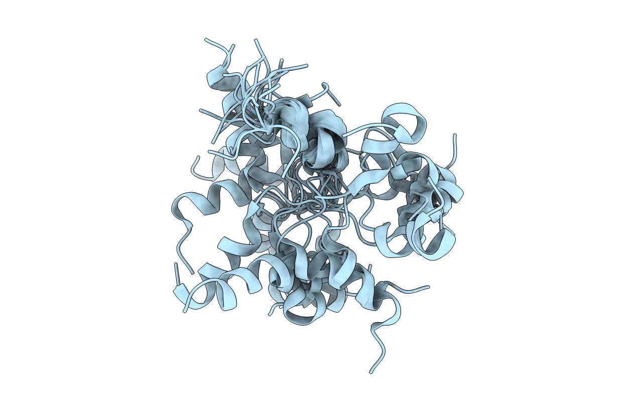
1QLO image
Deposition Date 1999-09-09
Release Date 1999-12-14
Last Version Date 2024-05-15
Entry Detail
PDB ID:
1QLO
Title:
Structure of the active domain of the herpes simplex virus protein ICP47 in water/sodium dodecyl sulfate solution determined by nuclear magnetic resonance spectroscopy
Biological Source:
Source Organism:
Host Organism:
Method Details:
Experimental Method:
Conformers Calculated:
40
Conformers Submitted:
19
Selection Criteria:
LEAST RESTRAINT VIOLATION ENERGY
Macromolecular Entities
Structures with similar UniProt ID
Protein Blast
Polymer Type:polypeptide(L)
Molecule:HERPES SIMPLEX VIRUS PROTEIN ICP47
Gene (Uniprot):US12
Chain IDs:A
Chain Length:34
Number of Molecules:1
Biological Source:HERPES SIMPLEX VIRUS
Ligand Molecules
Primary Citation
The Structure of the Active Domain of the Herpes Simplex Virus Protein Icp47 in Water/Sodium Dodecyl Sulfate Solution Determined by Nuclear Magnetic Resonance Spectroscopy.
Biochemistry 38 13692 ? (1999)
PMID: 10521276 DOI: 10.1021/BI9909647

Abstact

ICP47 encoded by herpes simplex virus (HSV) is a key factor in the evasion of cellular immune response against HSV-infected cells. By specific inhibition of the transporter associated with antigen processing (TAP), ICP47 prevents peptide transport into the endoplasmic reticulum and subsequent loading of major histocompatibility complex (MHC) class I molecules. Amino acid residues 3-34 have been identified as the active domain. This domain appeared to be unstructured in aqueous solution, whereas after binding to membranes an alpha-helical conformation was observed. Here, we have analyzed the structure of ICP47(2-34) in a lipidlike environment by nuclear magnetic resonance (NMR) spectroscopy. In micellar solution of deuterated sodium dodecyl sulfate, the viral TAP inhibitor adopts an ordered structure. There are two helical regions extending from residues 4 to 15 and from residues 22 to 32. Arg-16 is found on the C-terminus of the first helix, and Gly-33 serves as a terminator of the second helix. A loop between residues 17 and 21 is also evident in the structure. The relative orientation of the helices toward each other, however, could not be determined due to the paucity of NOEs from residues 18-21.

Legend

Protein

Chemical

Disease

Primary Citation of related structures