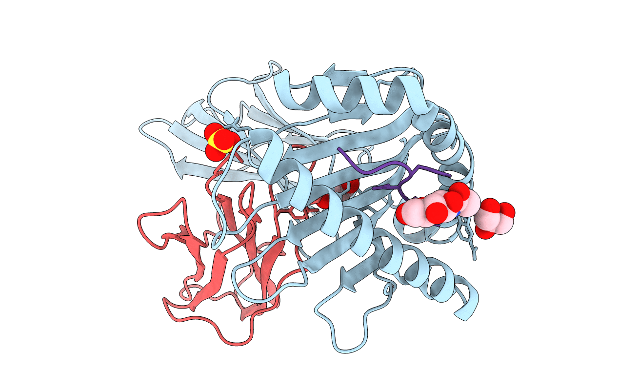
1QLF image
Entry Detail
PDB ID:
1QLF
Title:
MHC CLASS I H-2DB COMPLEXED WITH GLYCOPEPTIDE K3G
Biological Source:
Host Organism:
PDB Version:
Deposition Date:
1999-08-30
Release Date:
1999-09-01
Method Details:
Experimental Method:
Resolution:
2.65 Å
R-Value Free:
0.26
R-Value Work:
0.20
R-Value Observed:
0.20
Space Group:
P 1 21 1
Macromolecular Entities
Polymer Type:polypeptide(L)
Description:MHC CLASS I H-2DB HEAVY CHAIN
Chain IDs:A
Chain Length:276
Number of Molecules:1
Biological Source:MUS MUSCULUS
Polymer Type:polypeptide(L)
Description:HUMAN BETA-2-MICROGLOBULIN
Chain IDs:B
Chain Length:99
Number of Molecules:1
Biological Source:HOMO SAPIENS
Polymer Type:polypeptide(L)
Description:SYNTHETIC GLYCOPEPTIDE
Chain IDs:C
Chain Length:9
Number of Molecules:1
Biological Source:SENDAI VIRUS
Modified Residue
Compound ID Chain ID Parent Comp ID Details 2D Image
SER C SER GLYCOSYLATION SITE
Primary Citation
Crystal Structures of Two H-2Db/Glycopeptide Complexes Suggest a Molecular Basis for Ctl Cross-Reactivity
Immunity 10 63 ? (1999)
PMID: 10023771 DOI: 10.1016/S1074-7613(00)80007-2

Abstact

Two synthetic O-GlcNAc-bearing peptides that elicit H-2Db-restricted glycopeptide-specific cytotoxic T cells (CTL) have been shown to display nonreciprocal patterns of cross-reactivity. Here, we present the crystal structures of the H-2Db glycopeptide complexes to 2.85 A resolution or better. In both cases, the glycan is solvent exposed and available for direct recognition by the T cell receptor (TCR). We have modeled the complex formed between the MHC-glycopeptide complexes and their respective TCRs, showing that a single saccharide residue can be accommodated in the standard TCR-MHC geometry. The models also reveal a possible molecular basis for the observed cross-reactivity patterns of the CTL clones, which appear to be influenced by the length of the CDR3 loop and the nature of the immunizing ligand.

Legend

Protein

Chemical

Disease

Primary Citation of related structures