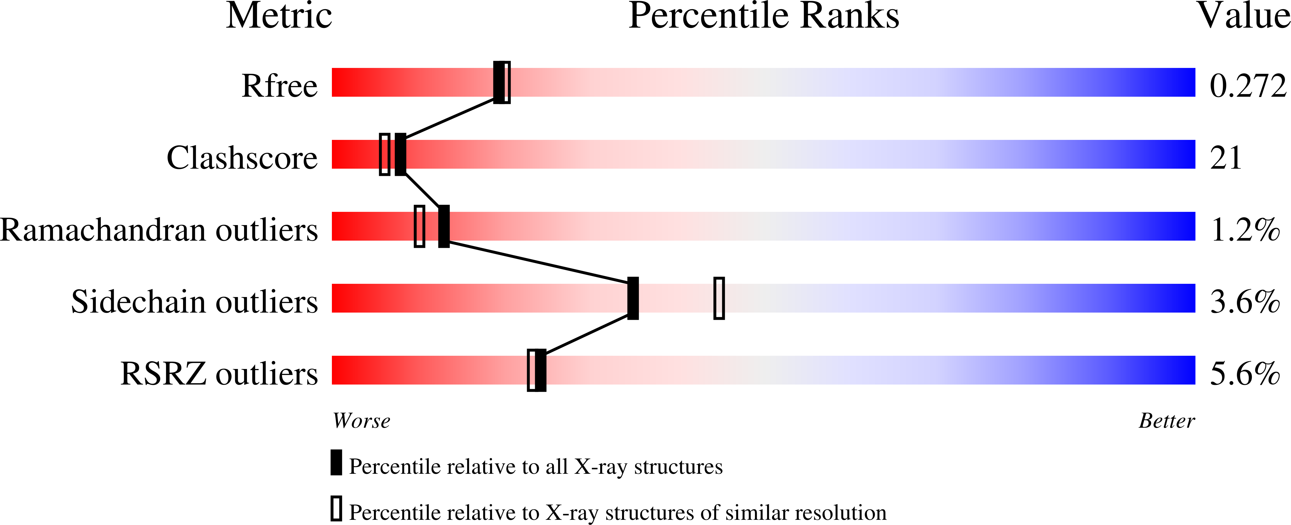
1QKT image
Deposition Date 1999-08-05
Release Date 2000-08-18
Last Version Date 2024-06-19
Entry Detail
PDB ID:
1QKT
Title:
MUTANT ESTROGEN NUCLEAR RECEPTOR LIGAND BINDING DOMAIN COMPLEXED WITH ESTRADIOL
Biological Source:
Source Organism:
HOMO SAPIENS (Taxon ID: 9606)
Host Organism:
Method Details:
Experimental Method:
Resolution:
2.20 Å
R-Value Free:
0.27
R-Value Work:
0.22
R-Value Observed:
0.22
Space Group:
P 65 2 2
Macromolecular Entities
Polymer Type:polypeptide(L)
Molecule:ESTRADIOL RECEPTOR
Gene (Uniprot):ESR1
Mutations:YES
Chain IDs:A
Chain Length:248
Number of Molecules:1
Biological Source:HOMO SAPIENS
Ligand Molecules
Primary Citation
Crystal Structure of a Mutant Heralpha Ligand- Binding Domain Reveals Key Structural Features for the Mechanism of Partial Agonism
J.Biol.Chem. 276 15059 ? (2001)
PMID: 11278577 DOI: 10.1074/JBC.M009870200

Abstact

The crystal structure of a triple cysteine to serine mutant ERalpha ligand-binding domain (LBD), complexed with estradiol, shows that despite the presence of a tightly bound agonist ligand, the protein exhibits an antagonist-like conformation, similar to that observed in raloxifen and 4-hydroxytamoxifen-bound structures. This mutated receptor binds estradiol with wild type affinity and displays transcriptional activity upon estradiol stimulation, but with limited potency (about 50%). This partial activity is efficiently repressed in antagonist competition assays. The comparison with available LBD structures reveals key features governing the positioning of helix H12 and highlights the importance of cysteine residues in promoting an active conformation. Furthermore the present study reveals a hydrogen bond network connecting ligand binding to protein trans conformation. These observations support a dynamic view of H12 positioning, where the control of the equilibrium between two stable locations determines the partial agonist character of a given ligand.

Legend

Protein

Chemical

Disease

Primary Citation of related structures