1QK4 image
Deposition Date 1999-07-09
Release Date 1999-10-17
Last Version Date 2023-12-13
Entry Detail
PDB ID:
1QK4
Keywords:
Title:
TOXOPLASMA GONDII HYPOXANTHINE-GUANINE PHOSPHORIBOSYLTRANSFERASE IMP COMPLEX
Biological Source:
Source Organism:
Host Organism:
Method Details:
Experimental Method:
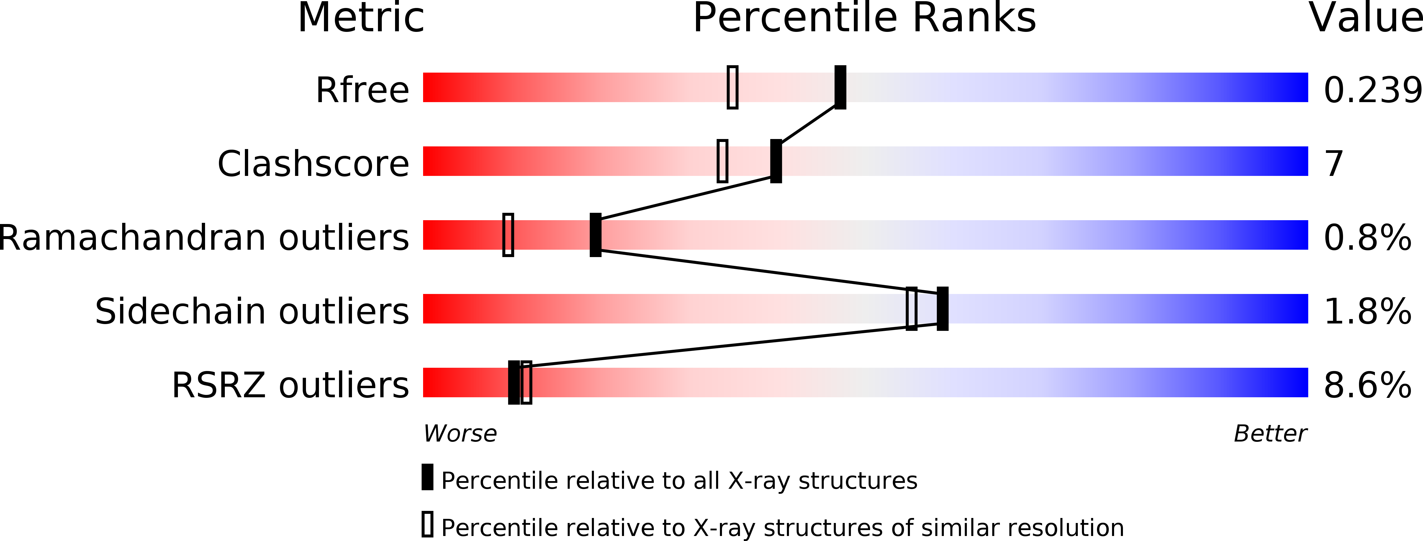
Resolution:
1.90 Å
R-Value Free:
0.23
R-Value Work:
0.18
Space Group:
P 21 21 21
Macromolecular Entities
Polymer Type:polypeptide(L)
Molecule:HYPOXANTHINE-GUANINE PHOSPHORIBOSYLTRANSFERASE
Gene (Uniprot):HPRT
Chain IDs:A, B, C, D
Chain Length:233
Number of Molecules:4
Biological Source:TOXOPLASMA GONDII
Ligand Molecules
Primary Citation
Crystal Structures of the Toxoplasma Gondii Hypoxanthine-Guanine Phosphoribosyltransferase Gmp and -Imp Complexes: Comparison of Purine Binding Interactions with the Xmp Complex
Biochemistry 38 14485 ? (1999)
PMID: 10545170 DOI: 10.1021/BI990507Q

Abstact

The crystal structures of the guanosine 5'-monophosphate (GMP) and inosine 5'-monophosphate (IMP) complexes of Toxoplasma gondii hypoxanthine-guanine phosphoribosyltransferase (HGPRT) have been determined at 1.65 and 1.90 A resolution. These complexes, which crystallize in space groups P2(1) (a = 65.45 A, b = 90.84 A, c = 80. 26 A, and beta = 92.53 degrees ) and P2(1)2(1)2(1) (a = 84.54 A, b = 102.44 A, and c = 108.83 A), each comprise a tetramer in the crystallographic asymmetric unit. All active sites in the tetramers are fully occupied by the nucleotide. Comparison of these structures with that of the xanthosine 5'-monophosphate (XMP)-pyrophosphate-Mg(2+) ternary complex reported in the following article [Heroux, A., et al. (1999) Biochemistry 38, 14495-14506] shows how T. gondii HGPRT is able to recognize guanine, hypoxanthine, and xanthine as substrates, and suggests why the human enzyme cannot use xanthine efficiently. Comparison with the apoenzyme reveals the structural changes that occur upon binding of purines and ribose 5'-phosphate to HGPRT. Two structural features important to the HGPRT mechanism, a previously unrecognized active site loop (loop III', residues 180-184) and an active site peptide bond (Leu78-Lys79) that adopts both the cis and the trans configurations, are presented.

Legend

Protein

Chemical

Disease

Primary Citation of related structures