1QK2 image
Entry Detail
PDB ID:
1QK2
Keywords:
Title:
WILD TYPE CEL6A WITH A NON-HYDROLYSABLE CELLOTETRAOSE
Biological Source:
Source Organism:
Host Organism:
PDB Version:
Deposition Date:
1999-07-09
Release Date:
1999-09-18
Method Details:
Experimental Method:
Resolution:
2.00 Å
R-Value Free:
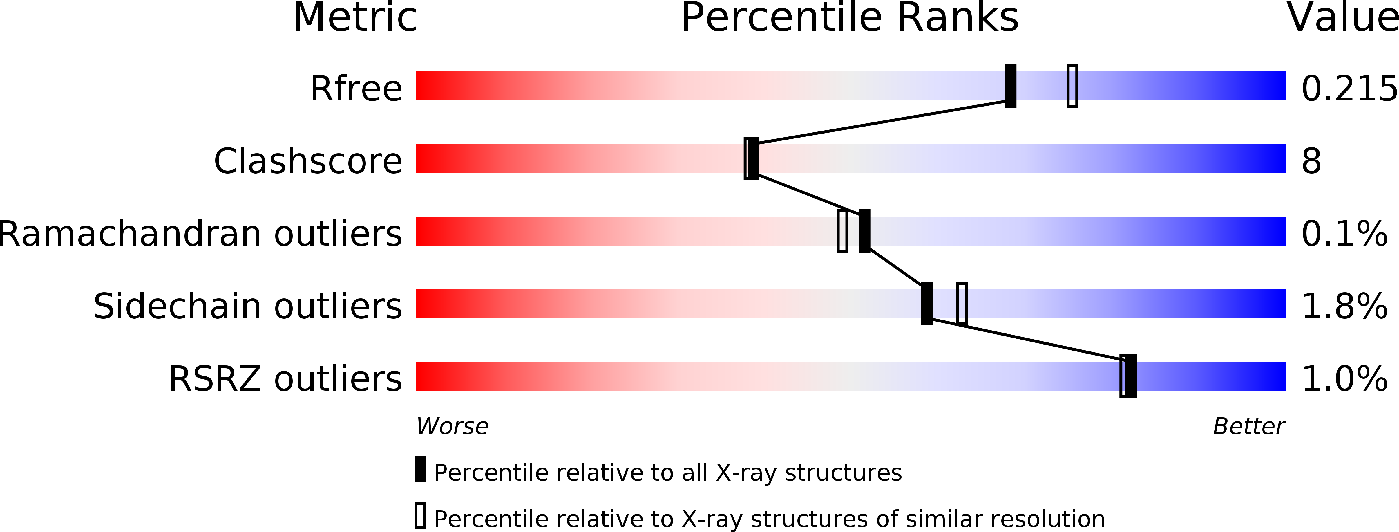
0.22
R-Value Work:
0.18
R-Value Observed:
0.18
Space Group:
P 1 21 1
Macromolecular Entities
Polymer Type:polypeptide(L)
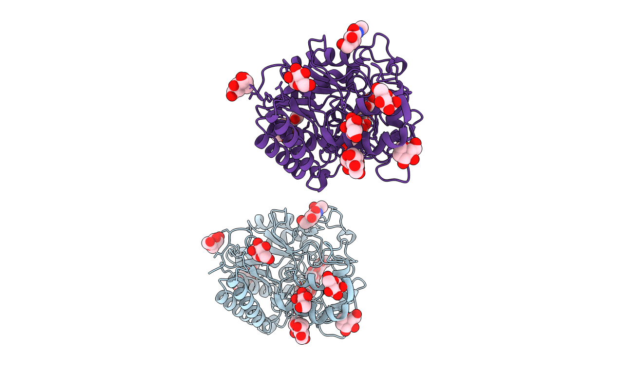
Description:CELLOBIOHYDROLASE CEL6A (FORMERLY CALLED CBH II)
Chain IDs:A, B
Chain Length:363
Number of Molecules:2
Biological Source:TRICHODERMA REESEI
Modified Residue
Compound ID Chain ID Parent Comp ID Details 2D Image
ASN A ASN GLYCOSYLATION SITE
SER A SER GLYCOSYLATION SITE
THR A THR GLYCOSYLATION SITE
Primary Citation
Crystallographic Evidence for Substrate Ring Distortion and Protein Conformational Changes During Catalysis in Cellobiohydrolase Cel6A from Trichoderma Reesei
Structure 7 1035 ? (1999)
PMID: 10508787 DOI: 10.1016/S0969-2126(99)80171-3

Abstact

BACKGROUND: Cel6A is one of the two cellobiohydrolases produced by Trichoderma reesei. The catalytic core has a structure that is a variation of the classic TIM barrel. The active site is located inside a tunnel, the roof of which is formed mainly by a pair of loops. RESULTS: We describe three new ligand complexes. One is the structure of the wild-type enzyme in complex with a nonhydrolysable cello-oligosaccharide, methyl 4-S-beta-cellobiosyl-4-thio-beta-cellobioside (Glc)(2)-S-(Glc)(2), which differs from a cellotetraose in the nature of the central glycosidic linkage where a sulphur atom replaces an oxygen atom. The second structure is a mutant, Y169F, in complex with the same ligand, and the third is the wild-type enzyme in complex with m-iodobenzyl beta-D-glucopyranosyl-beta(1,4)-D-xylopyranoside (IBXG). CONCLUSIONS: The (Glc)(2)-S-(Glc)(2) ligand binds in the -2 to +2 sites in both the wild-type and mutant enzymes. The glucosyl unit in the -1 site is distorted from the usual chair conformation in both structures. The IBXG ligand binds in the -2 to +1 sites, with the xylosyl unit in the -1 site where it adopts the energetically favourable chair conformation. The -1 site glucosyl of the (Glc)(2)-S-(Glc)(2) ligand is unable to take on this conformation because of steric clashes with the protein. The crystallographic results show that one of the tunnel-forming loops in Cel6A is sensitive to modifications at the active site, and is able to take on a number of different conformations. One of the conformational changes disrupts a set of interactions at the active site that we propose is an integral part of the reaction mechanism.

Legend

Protein

Chemical

Disease

Primary Citation of related structures