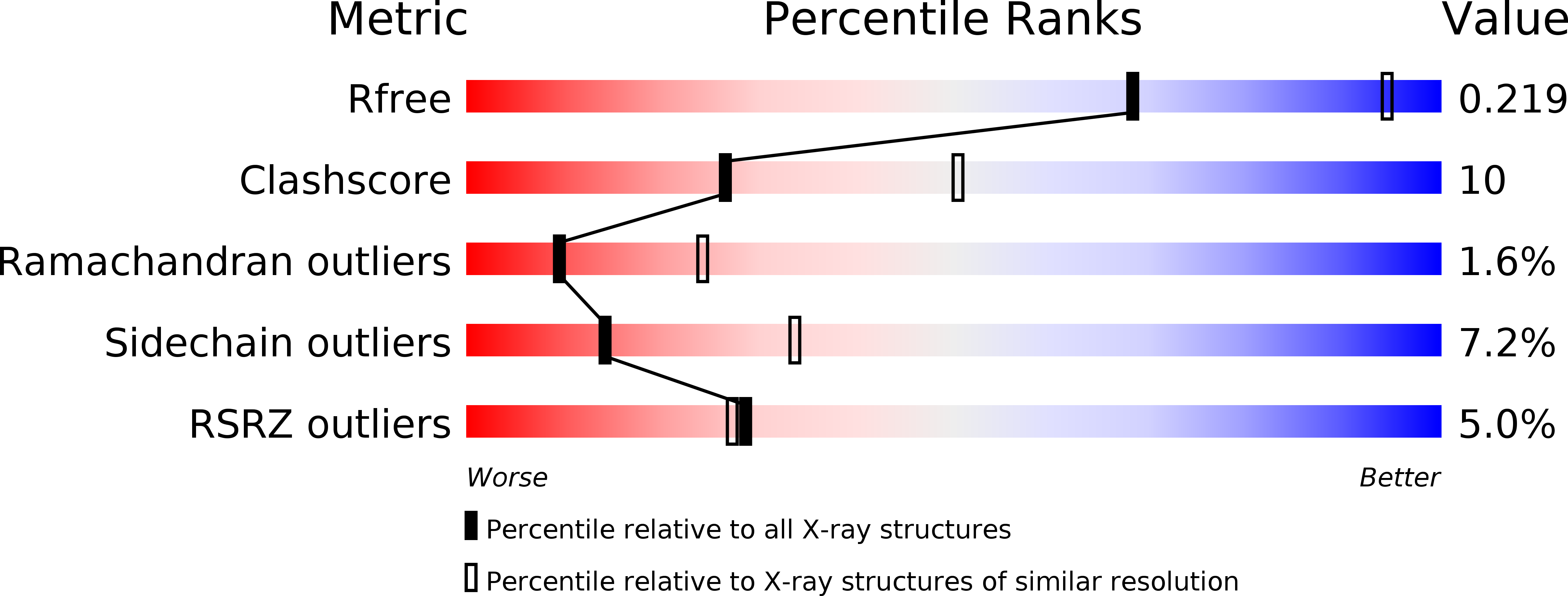
1QK1 image
Deposition Date 1999-07-08
Release Date 2000-04-11
Last Version Date 2023-12-13
Entry Detail
PDB ID:
1QK1
Title:
CRYSTAL STRUCTURE OF HUMAN UBIQUITOUS MITOCHONDRIAL CREATINE KINASE
Biological Source:
Source Organism:
HOMO SAPIENS (Taxon ID: 9606)
Method Details:
Experimental Method:
Resolution:
2.70 Å
R-Value Free:
0.21
R-Value Work:
0.19
R-Value Observed:
0.19
Space Group:
P 1 21 1
Macromolecular Entities
Polymer Type:polypeptide(L)
Molecule:CREATINE KINASE, UBIQUITOUS MITOCHONDRIAL
Gene (Uniprot):CKMT1A, CKMT1B
Chain IDs:A, B, C, D, E, F, G, H
Chain Length:379
Number of Molecules:8
Biological Source:HOMO SAPIENS
Ligand Molecules
Primary Citation
Crystal Structure of Human Ubiquitous Mitochondrial Creatine Kinase
Proteins: Struct.,Funct., Genet. 39 216 ? (2000)
PMID: 10737943 DOI: 10.1002/(SICI)1097-0134(20000515)39:3<216::AID-PROT40>3.0.CO;2-#

Abstact

Creatine kinase (CK), catalyzing the reversible trans-phosphorylation between ATP and creatine, plays a key role in the energy metabolism of cells with high and fluctuating energy requirements. We have solved the X-ray structure of octameric human ubiquitous mitochondrial CK (uMtCK) at 2.7 A resolution, representing the first human CK structure. The structure is very similar to the previously determined structure of sarcomeric mitochondrial CK (sMtCK). The cuboidal octamer has 422 point group symmetry with four dimers arranged along the fourfold axis and a central channel of approximately 20 A diameter, which extends through the whole octamer. Structural differences with respect to sMtCK are found in isoform-specific regions important for octamer formation and membrane binding. Octameric uMtCK is stabilized by numerous additional polar interactions between the N-termini of neighboring dimers, which extend into the central channel and form clamp-like structures, and by a pair of salt bridges in the hydrophobic interaction patch. The five C-terminal residues of uMtCK, carrying positive charges likely to be involved in phospholipid-binding, are poorly defined by electron density, indicating a more flexible region than the corresponding one in sMtCK. The structural differences between uMtCK and sMtCK are consistent with biochemical studies on octamer stability and membrane binding of the two isoforms.

Legend

Protein

Chemical

Disease

Primary Citation of related structures