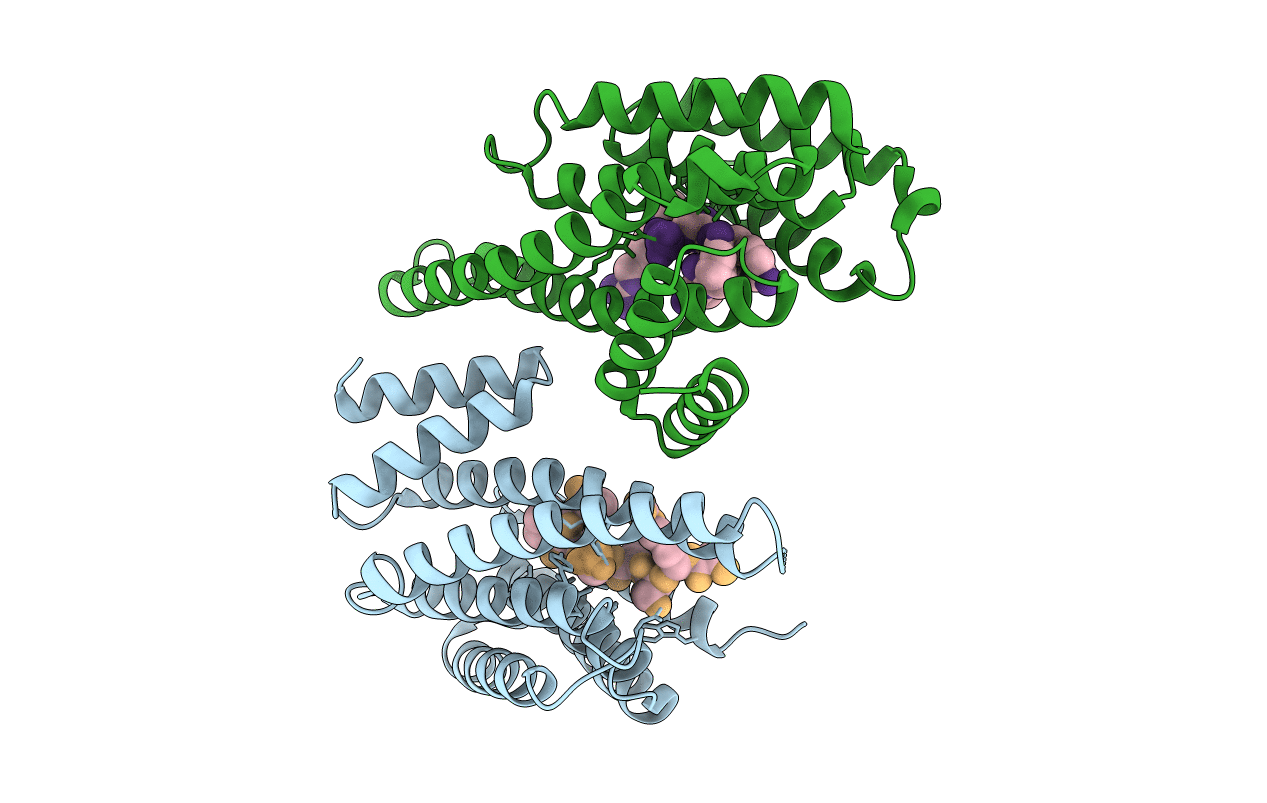
1QJB image
Deposition Date 1999-06-23
Release Date 1999-09-15
Last Version Date 2024-10-23
Entry Detail
PDB ID:
1QJB
Title:
14-3-3 ZETA/PHOSPHOPEPTIDE COMPLEX (MODE 1)
Biological Source:
Source Organism:
HOMO SAPIENS (Taxon ID: 9606)
Method Details:
Experimental Method:
Resolution:
2.00 Å
R-Value Free:
0.28
R-Value Work:
0.21
Space Group:
P 21 21 21
Macromolecular Entities
Polymer Type:polypeptide(L)
Molecule:14-3-3 PROTEIN ZETA/DELTA
Chain IDs:A, B
Chain Length:245
Number of Molecules:2
Biological Source:HOMO SAPIENS
Polymer Type:polypeptide(L)
Molecule:PHOSPHOPEPTIDE
Chain IDs:C (auth: Q), D (auth: S)
Chain Length:8
Number of Molecules:2
Biological Source:HOMO SAPIENS
Modified Residue
Compound ID Chain ID Parent Comp ID Details 2D Image
SEP C SER PHOSPHOSERINE
Primary Citation
Structural Analysis of 14-3-3 Phosphopeptide Complexes Identifies a Dual Role for the Nuclear Export Signal of 14-3-3 in Ligand Binding
Mol.Cell 4 153 ? (1999)
PMID: 10488331 DOI: 10.1016/S1097-2765(00)80363-9

Abstact

We have solved the high-resolution X-ray structure of 14-3-3 bound to two different phosphoserine peptides, representing alternative substrate-binding motifs. These structures reveal an evolutionarily conserved network of peptide-protein interactions within all 14-3-3 isotypes, explain both binding motifs, and identify a novel intrachain phosphorylation-mediated loop structure in one of the peptides. A 14-3-3 mutation disrupting Raf signaling alters the ligand-binding cleft, selecting a different phosphopeptide-binding motif and different substrates than the wild-type protein. Many 14-3-3: peptide contacts involve a C-terminal amphipathic alpha helix containing a putative nuclear export signal, implicating this segment in both ligand and Crm1 binding. Structural homology between the 14-3-3 NES structure and those within I kappa B alpha and p53 reveals a conserved topology recognized by the Crm1 nuclear export machinery.

Legend

Protein

Chemical

Disease

Primary Citation of related structures