1QJ8 image
Deposition Date 1999-06-23
Release Date 1999-10-10
Last Version Date 2024-05-08
Entry Detail
PDB ID:
1QJ8
Title:
CRYSTAL STRUCTURE OF THE OUTER MEMBRANE PROTEIN OMPX FROM ESCHERICHIA COLI
Biological Source:
Source Organism:
ESCHERICHIA COLI (Taxon ID: 562)
Host Organism:
Method Details:
Experimental Method:
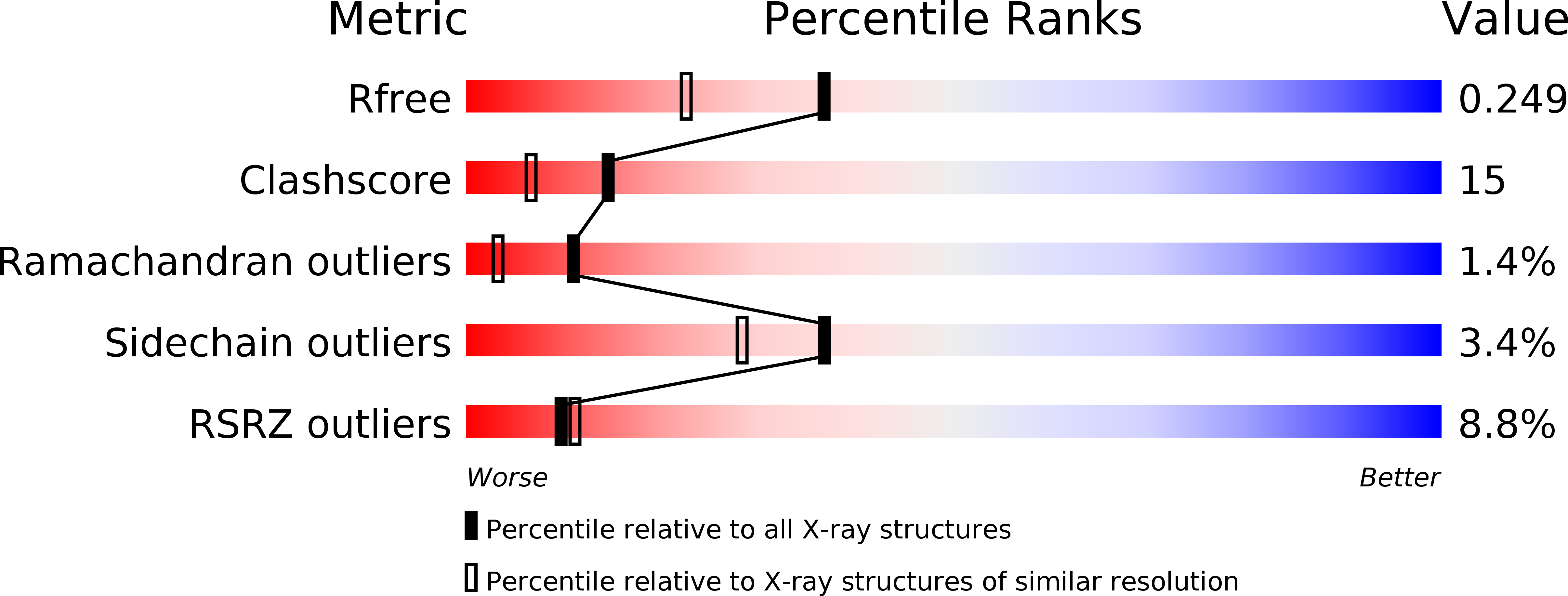
Resolution:
1.90 Å
R-Value Free:
0.24
R-Value Work:
0.20
Space Group:
H 3 2
Macromolecular Entities
Polymer Type:polypeptide(L)
Molecule:OUTER MEMBRANE PROTEIN X
Mutations:YES
Chain IDs:A
Chain Length:148
Number of Molecules:1
Biological Source:ESCHERICHIA COLI
Primary Citation
The Structure of the Outer Membrane Protein Ompx from Escherichia Coli Reveals Mechanisms of Virulence
Structure 7 1301 ? (1999)
PMID: 10545325 DOI: 10.1016/S0969-2126(00)80063-5

Abstact

BACKGROUND: The integral outer membrane protein X (OmpX) from Escherichia coli belongs to a family of highly conserved bacterial proteins that promote bacterial adhesion to and entry into mammalian cells. Moreover, these proteins have a role in the resistance against attack by the human complement system. Here we present the first crystal structure of a member of this family. RESULTS: The crystal structure of OmpX from E. coli was determined at 1.9 A resolution using multiple isomorphous replacement. OmpX consists of an eight-stranded antiparallel all-next-neighbor beta barrel. The structure shows two girdles of aromatic amino acid residues and a ribbon of nonpolar residues that attach to the membrane interior. The core of the barrel consists of an extended hydrogen-bonding network of highly conserved residues. OmpX thus resembles an inverse micelle. The structure explains the dramatically improved crystal quality of OmpX containing the mutation His100-->Asn, which made the X-ray analysis possible. The coordination spheres of two bound platinum ions are described. CONCLUSIONS: The OmpX structure shows that within a family of virulence-related membrane proteins, the membrane-spanning part of the protein is much better conserved than the extracellular loops. Moreover, these loops form a protruding beta sheet, the edge of which presumably binds to external proteins. It is suggested that this type of binding promotes cell adhesion and invasion and helps defend against the complement system. Although OmpX has the same beta-sheet topology as the structurally related outer membrane protein A (OmpA), their barrels differ with respect to the shear numbers and internal hydrogen-bonding networks.

Legend

Protein

Chemical

Disease

Primary Citation of related structures