Deposition Date
1999-06-02
Release Date
2000-06-07
Last Version Date
2023-12-27
Entry Detail
PDB ID:
1QI0
Keywords:
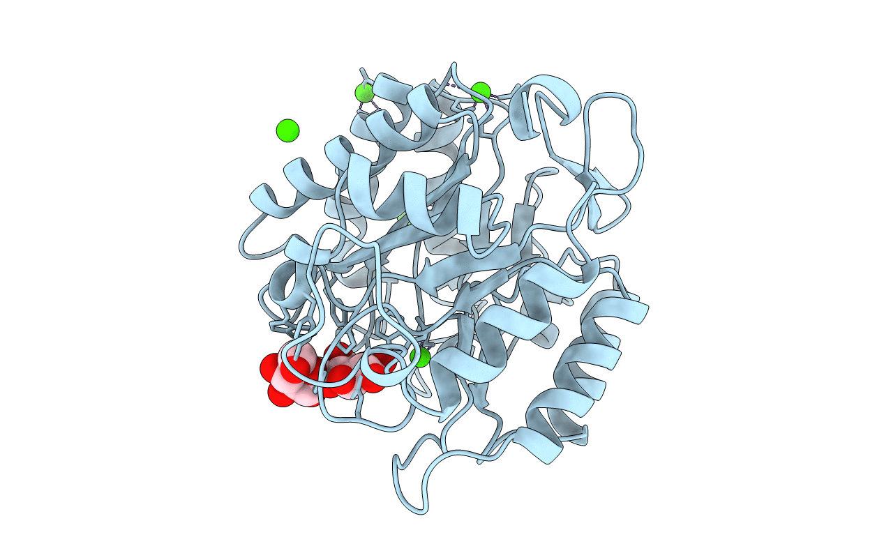
Title:
ENDOGLUCANASE CEL5A FROM BACILLUS AGARADHAERENS IN THE TETRAGONAL CRYSTAL FORM IN COMPLEX WITH CELLOBIOSE
Biological Source:
Source Organism:
Bacillus agaradhaerens (Taxon ID: 76935)
Host Organism:
Method Details:
Experimental Method:
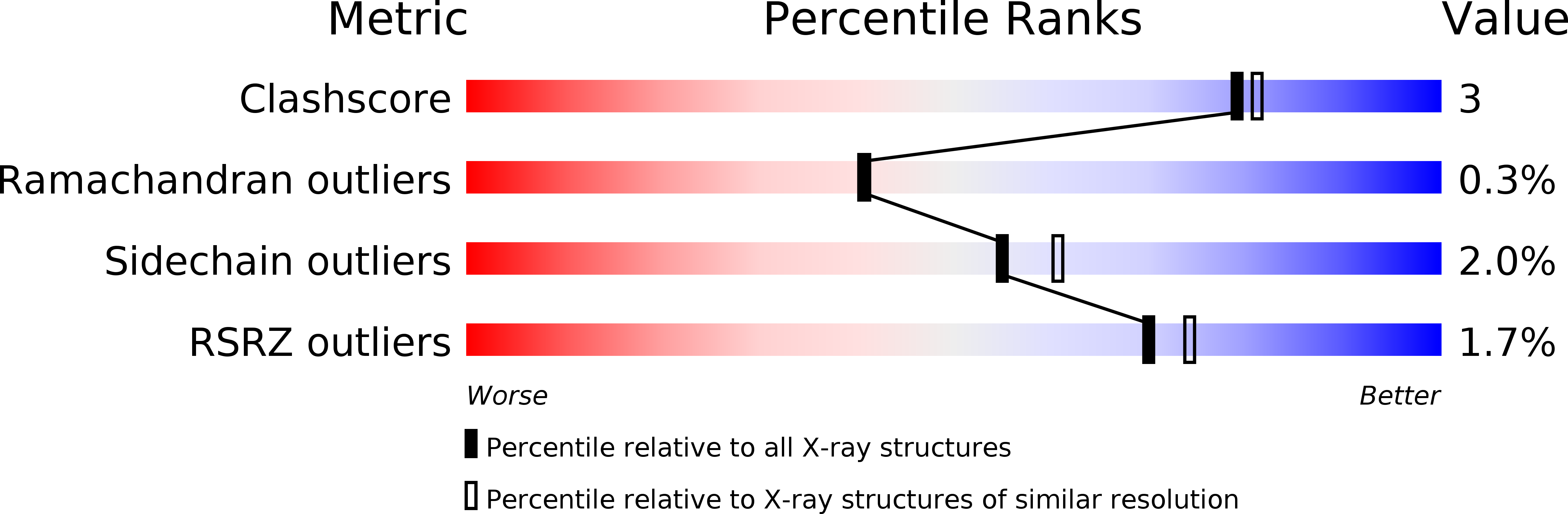
Resolution:
2.10 Å
R-Value Free:
0.21
R-Value Work:
0.17
R-Value Observed:
0.16
Space Group:
P 43 21 2