
Deposition Date
1999-05-11
Release Date
1999-11-10
Last Version Date
2023-08-16
Entry Detail
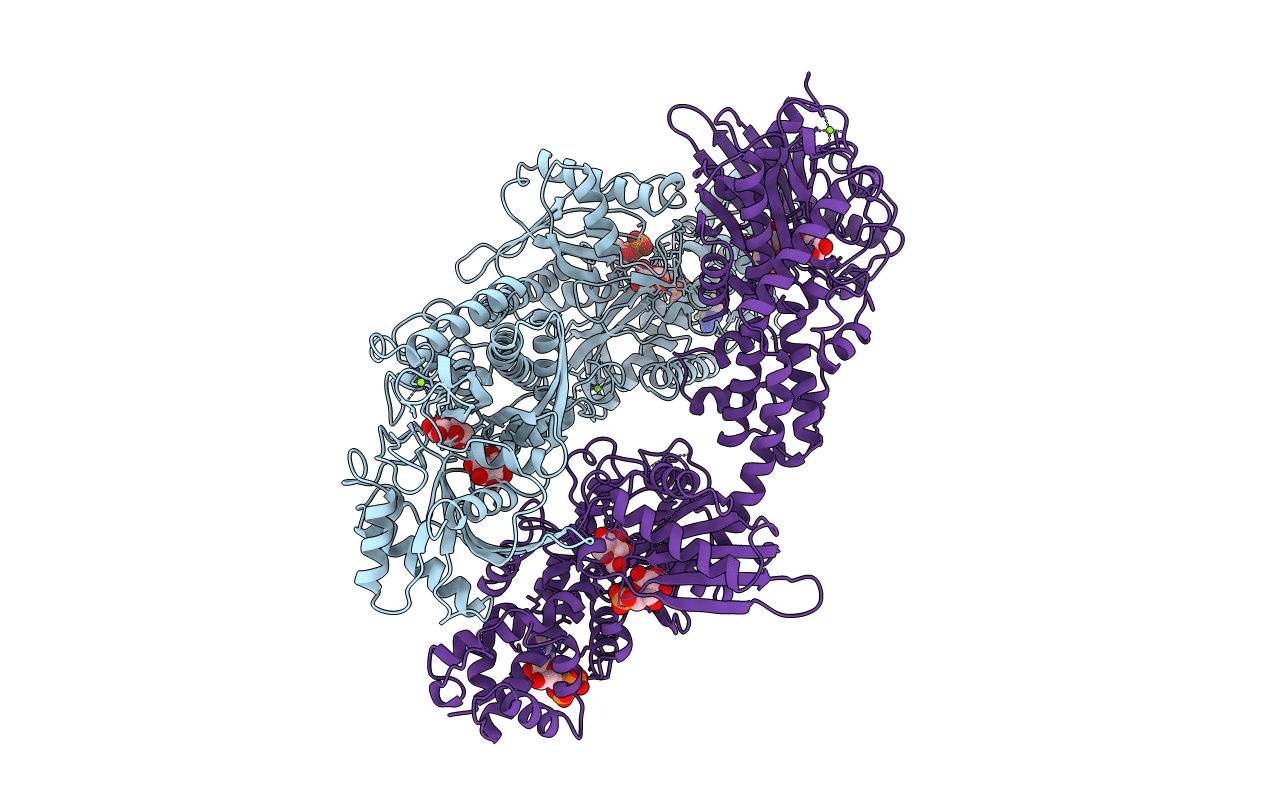
PDB ID:
1QHA
Keywords:
Title:
HUMAN HEXOKINASE TYPE I COMPLEXED WITH ATP ANALOGUE AMP-PNP
Biological Source:
Source Organism(s):
Homo sapiens (Taxon ID: 9606)
Expression System(s):
Method Details:
Experimental Method:
Resolution:
2.25 Å
R-Value Free:
0.27
R-Value Work:
0.20
Space Group:
P 1 21 1
