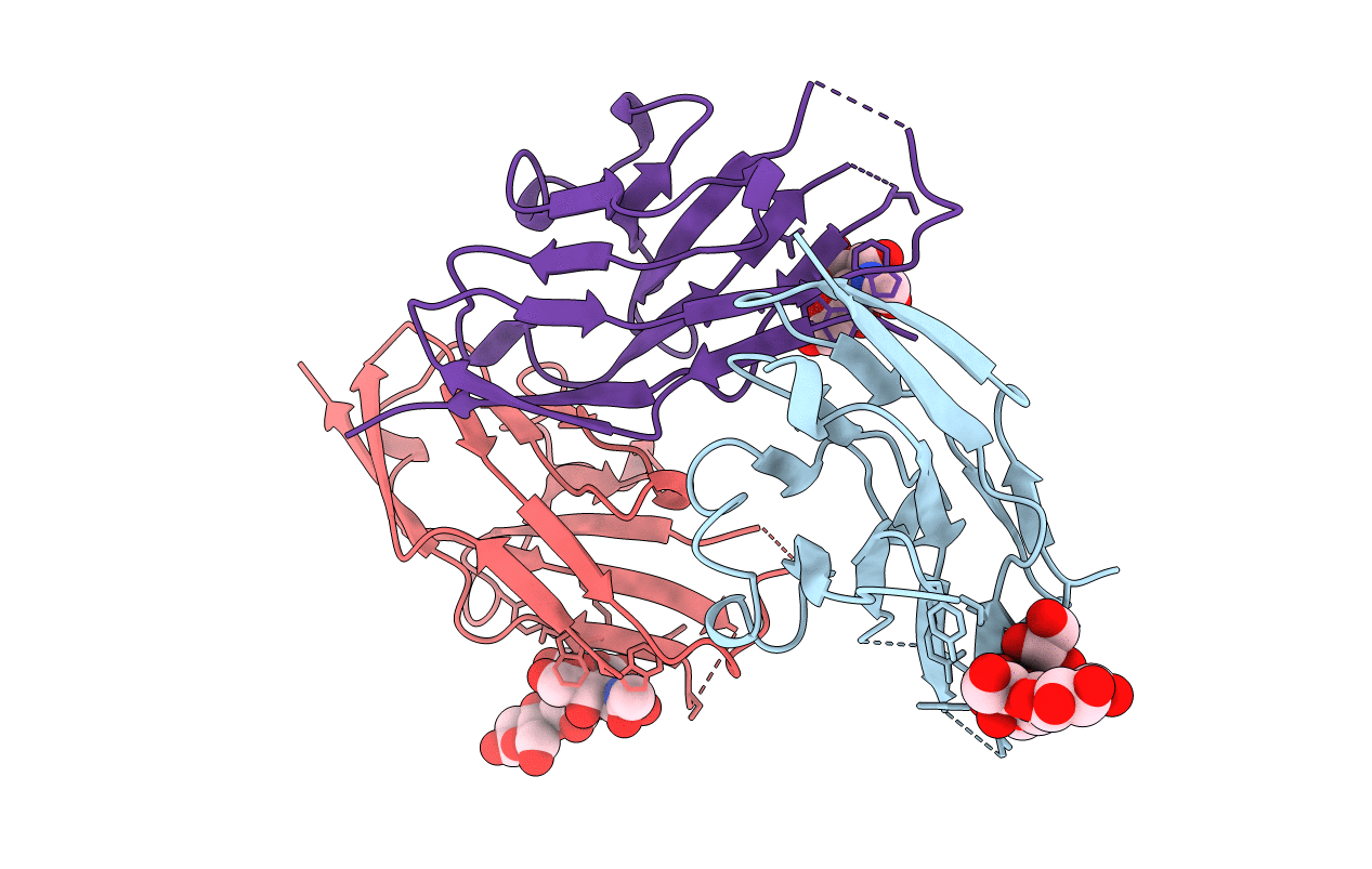
1QFO image
Deposition Date 1999-04-12
Release Date 1999-04-16
Last Version Date 2024-10-30
Entry Detail
PDB ID:
1QFO
Keywords:
Title:
N-TERMINAL DOMAIN OF SIALOADHESIN (MOUSE) IN COMPLEX WITH 3'SIALYLLACTOSE
Biological Source:
Source Organism:
Mus musculus (Taxon ID: 10090)
Host Organism:
Method Details:
Experimental Method:
Resolution:
1.85 Å
R-Value Free:
0.23
R-Value Work:
0.2
Space Group:
P 21 21 21
Macromolecular Entities
Polymer Type:polypeptide(L)
Molecule:PROTEIN (SIALOADHESIN)
Chain IDs:A, B, C
Chain Length:119
Number of Molecules:3
Biological Source:Mus musculus
Ligand Molecules
Peptide-like Molecules
PRD_900025
Primary Citation
Crystal structure of the N-terminal domain of sialoadhesin in complex with 3' sialyllactose at 1.85 A resolution.
Mol.Cell 1 719 728 (1998)
PMID: 9660955 DOI: 10.1016/S1097-2765(00)80071-4

Abstact

The structure of the functional N-terminal domain from the extracellular region of the cell surface receptor sialoadhesin has been determined in complex with the oligosaccharide 3' sialyllactose. This provides structural information for the siglec family of proteins. The structure conforms to the V-set immunoglobulin-like fold but contains several distinctive features, including an intra-beta sheet disulphide and a splitting of the standard beta strand G into two shorter strands. These novel features appear important in adapting the V-set fold for sialic acid-mediated recognition. Analysis of the complex with 3'sialyllactose highlights three residues, conserved throughout the siglec family, as key features of the sialic acid-binding template. The complex is representative of the functional recognition interaction with carbohydrate and as such provides detailed information for a heterotypic cell adhesion interaction.

Legend

Protein

Chemical

Disease

Primary Citation of related structures