1QFL image
Deposition Date 1999-04-12
Release Date 2000-04-19
Last Version Date 2024-10-30
Entry Detail
PDB ID:
1QFL
Keywords:
Title:
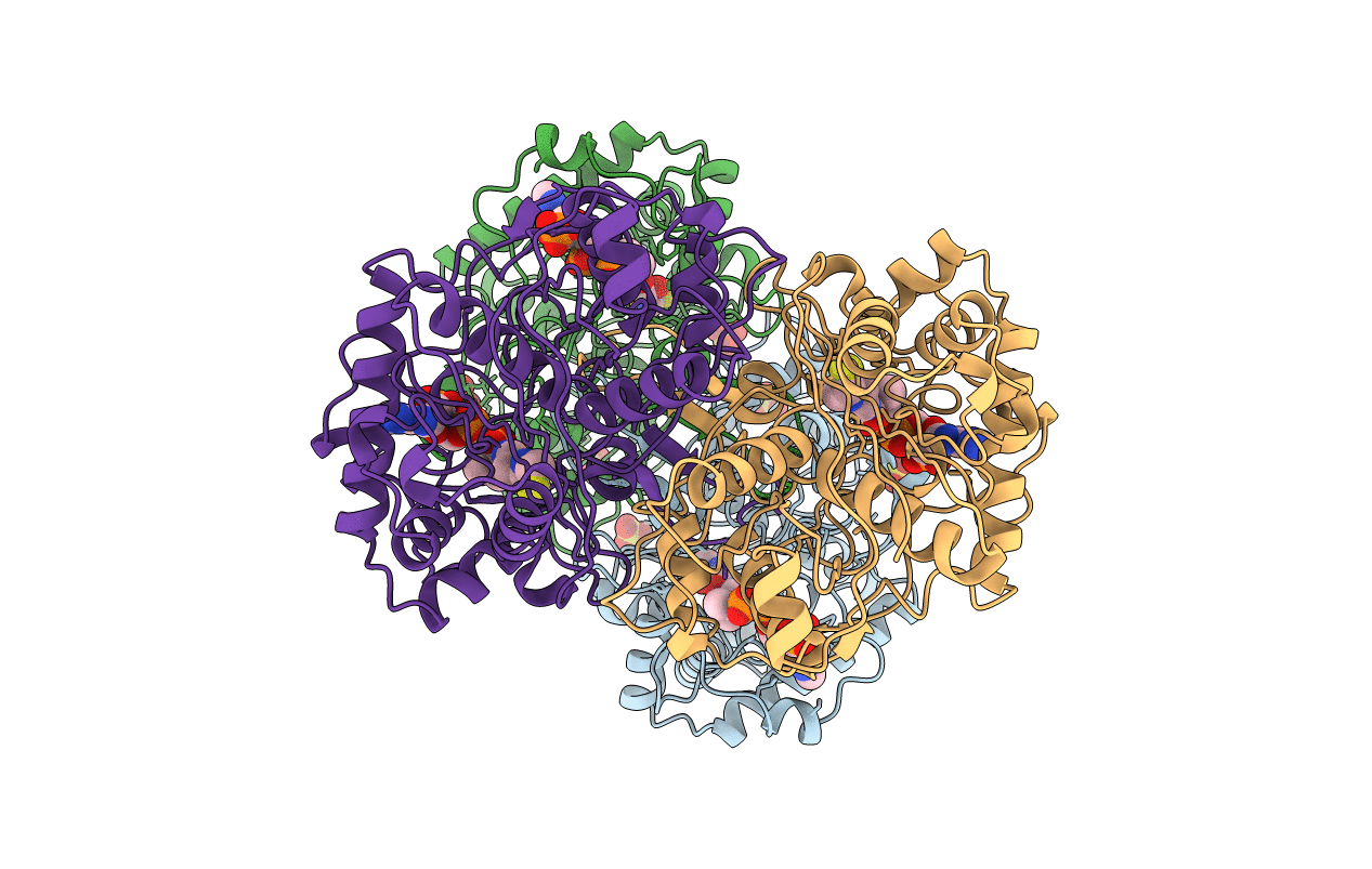
BIOSYNTHETIC THIOLASE FROM ZOOGLOEA RAMIGERA IN COMPLEX WITH A REACTION INTERMEDIATE.
Biological Source:
Source Organism:
Host Organism:
Method Details:
Experimental Method:
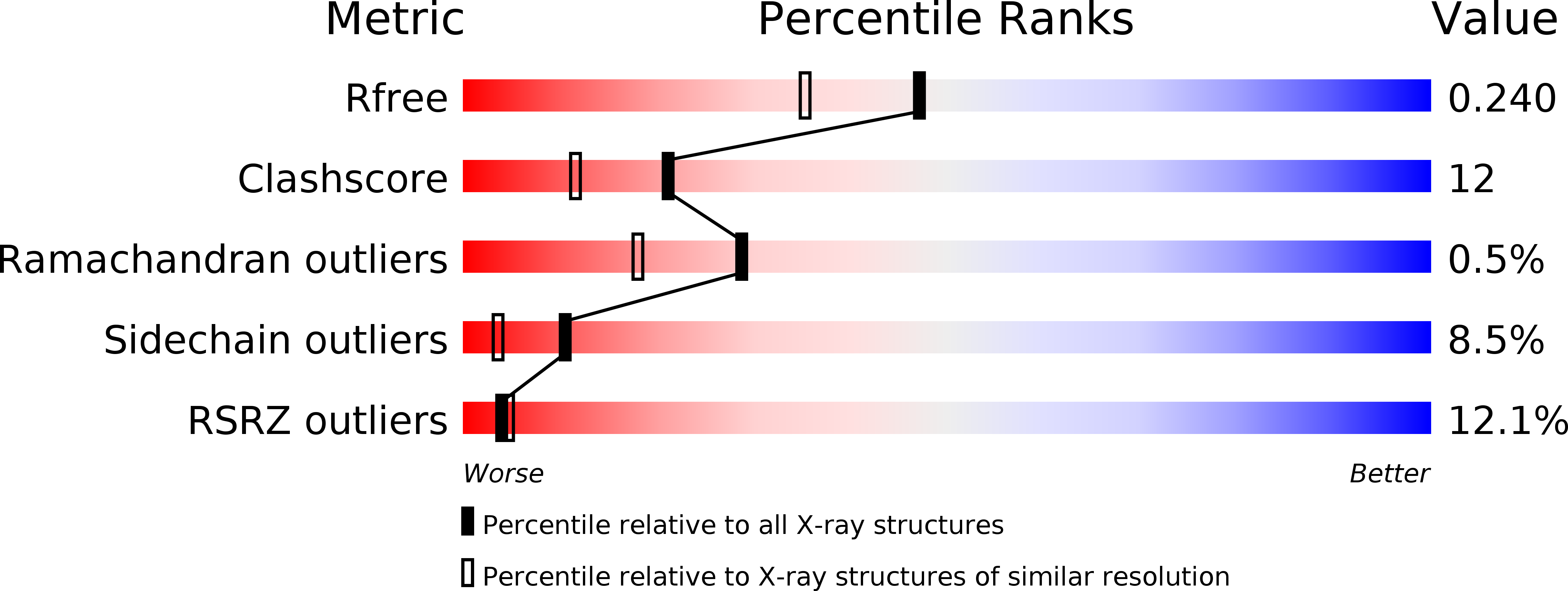
Resolution:
1.92 Å
R-Value Free:
0.25
R-Value Work:
0.20
Space Group:
P 1 21 1
Macromolecular Entities
Structures with similar UniProt ID
Protein Blast
Polymer Type:polypeptide(L)
Molecule:PROTEIN (ACETOACETYL-COA THIOLASE)
Gene (Uniprot):phaA
Chain IDs:A, B, C, D
Chain Length:389
Number of Molecules:4
Biological Source:Zoogloea ramigera
Modified Residue
Compound ID Chain ID Parent Comp ID Details 2D Image
SCY A CYS S-ACETYL-CYSTEINE
Primary Citation
A biosynthetic thiolase in complex with a reaction intermediate: the crystal structure provides new insights into the catalytic mechanism.
Structure Fold.Des. 7 1279 1290 (1999)
PMID: 10545327 DOI: 10.1016/S0969-2126(00)80061-1

Abstact

BACKGROUND: Thiolases are ubiquitous and form a large family of dimeric or tetrameric enzymes with a conserved, five-layered alphabetaalphabetaalpha catalytic domain. Thiolases can function either degradatively, in the beta-oxidation pathway of fatty acids, or biosynthetically. Biosynthetic thiolases catalyze the biological Claisen condensation of two molecules of acetyl-CoA to form acetoacetyl-CoA. This is one of the fundamental categories of carbon skeletal assembly patterns in biological systems and is the first step in a wide range of biosynthetic pathways, including those that generate cholesterol, steroid hormones, and various energy-storage molecules. RESULTS: The crystal structure of the tetrameric biosynthetic thiolase from Zoogloea ramigera has been determined at 2.0 A resolution. The structure contains a striking and novel 'cage-like' tetramerization motif, which allows for some hinge motion of the two tight dimers with respect to each other. The protein crystals were flash-frozen after a short soak with the enzyme's substrate, acetoacetyl-CoA. A reaction intermediate was thus trapped: the enzyme tetramer is acetylated at Cys89 and has a CoA molecule bound in each of its active-site pockets. CONCLUSIONS: The shape of the substrate-binding pocket reveals the basis for the short-chain substrate specificity of the enzyme. The active-site architecture, and in particular the position of the covalently attached acetyl group, allow a more detailed reaction mechanism to be proposed in which Cys378 is involved in both steps of the reaction. The structure also suggests an important role for the thioester oxygen atom of the acetylated enzyme in catalysis.

Legend

Protein

Chemical

Disease

Primary Citation of related structures