1QCY image
Deposition Date 1999-05-12
Release Date 2003-09-02
Last Version Date 2024-02-14
Entry Detail
PDB ID:
1QCY
Keywords:
Title:
THE CRYSTAL STRUCTURE OF THE I-DOMAIN OF HUMAN INTEGRIN ALPHA1BETA1
Biological Source:
Source Organism:
Homo sapiens (Taxon ID: 9606)
Host Organism:
Method Details:
Experimental Method:
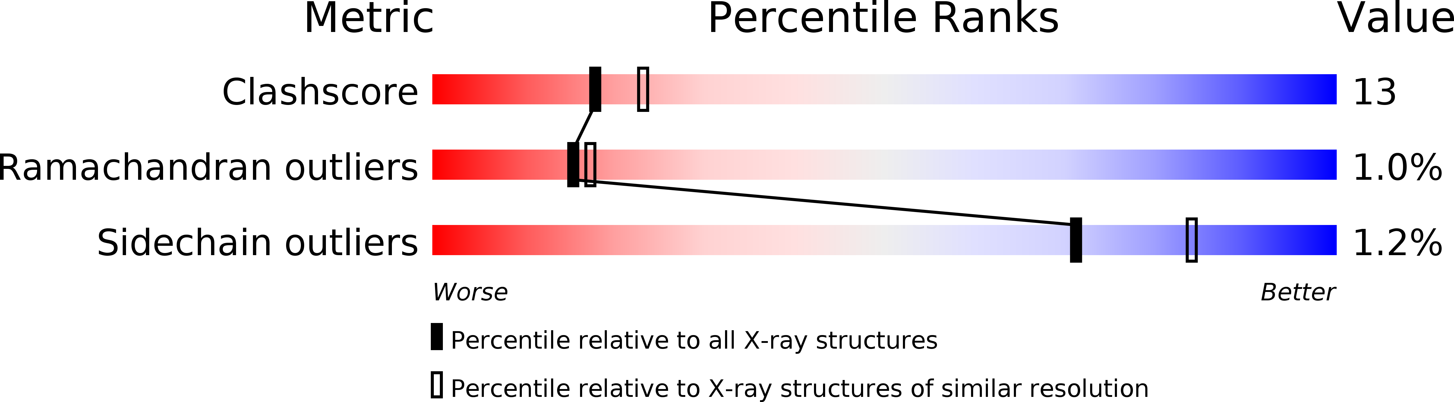
Resolution:
2.30 Å
R-Value Free:
0.25
R-Value Work:
0.2
Space Group:
C 1 2 1
Macromolecular Entities
Polymer Type:polypeptide(L)
Molecule:I-DOMAIN OF INTEGRIN ALPHA1BETA1
Gene (Uniprot):ITGA1
Mutations:K170E
Chain IDs:A
Chain Length:193
Number of Molecules:1
Biological Source:Homo sapiens
Ligand Molecules
Primary Citation
Jararhagin-derived RKKH Peptides Induce Structural Changes in a1I Domain of Human Integrin a1b1
J.Biol.Chem. 279 7962 7970 (2004)
PMID: 14660600 DOI: 10.1074/jbc.M312912200

Abstact

Integrin alpha(1)beta(1) is one of four collagen-binding integrins in humans. Collagens bind to the alphaI domain and in the case of alpha(2)I collagen binding is competitively inhibited by peptides containing the RKKH sequence and derived from the metalloproteinase jararhagin of snake venom from Bothrops jararaca. In alpha(2)I, these peptides bind near the metal ion-dependent adhesion site (MIDAS), where a collagen (I)-like peptide is known to bind; magnesium is required for binding. Published structures of the ligand-bound "open" conformation of alpha(2)I differs significantly from the "closed" conformation seen in the structure of apo-alpha(2)I near MIDAS. Here we show that two peptides, CTRKKHDC and CARKKHDC, derived from jararhagin also bind to alpha(1)I and competitively inhibit collagen I binding. Furthermore, calorimetric and fluorimetric measurements show that the structure of the complex of alpha(1)I with Mg(2+) and CTRKKHDC differs from structure in the absence of peptide. A comparison of the x-ray structure of apo-alpha(1)I ("closed" conformation) and a model structure of the alpha(1)I ("open" conformation) based on the closely related structure of alpha(2)I reveals that the binding site is partially blocked to ligands by Glu(255) and Tyr(285) in the "closed" structure, whereas in the "open" structure helix C is unwound and these residues are shifted, and the "RKKH" peptides fit well when docked. The "open" conformation of alpha(2)I resulting from binding a collagen (I)-like peptide leads to exposure of hydrophobic surface, also seen in the model of alpha(1)I and shown experimentally for alpha(1)I using a fluorescent hydrophobic probe.

Legend

Protein

Chemical

Disease

Primary Citation of related structures