Abstact
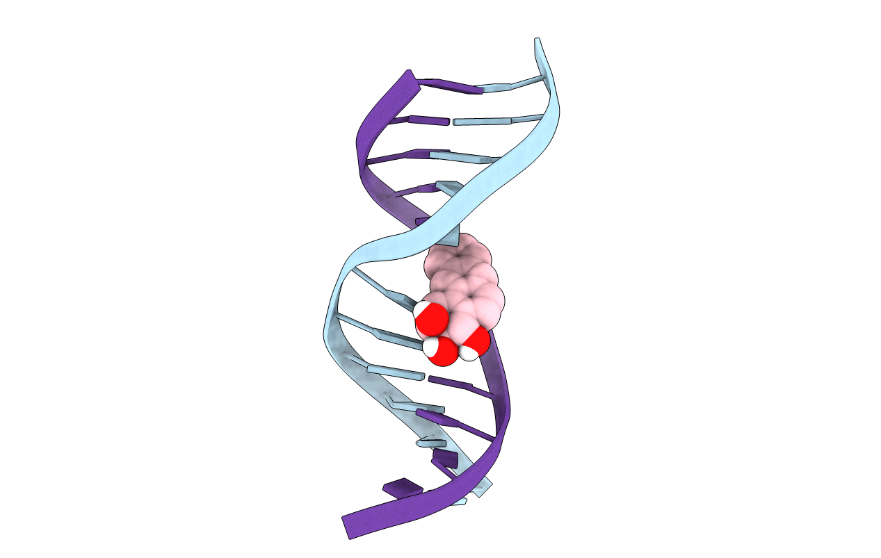
The solution structure of the (-)-(1R,2S,3R,4S)-N6-[1-(1,2,3, 4-tetrahydroxy-benz[a]anthracenyl)]-2'-deoxyadenosyl adduct at X6 of 5'-d(CGGACXAGAAG)-3'.5'-d(CTTCTTGTCCG)-3', incorporating codons 60, 61(italic), and 62 of the human N-ras protooncogene, was determined. This adduct results from the trans opening of 1S,2R,3R,4S-1, 2-epoxy-1,2,3,4-tetrahydro-benz[a]anthracenyl-3,4-diol by the exocyclic N6 of adenine. Molecular dynamics simulations were restrained by 509 NOEs from 1H NMR. The precision of the refined structures was monitored by pairwise root-mean-square deviations which were <1.2 A; accuracy was measured by complete relaxation matrix calculations, which yielded a sixth root R factor of 9.1 x 10(-)2 at 250 ms. The refined structure was a right-handed duplex, in which the benz[a]anthracene moiety intercalated from the major groove between C5.G18 and R,S,R,SA6.T17. In this orientation, the saturated ring of BA was oriented in the major groove of the duplex, with the aromatic rings inserted into the duplex such that the terminal ring of BA threaded the duplex and faced toward the minor groove direction. The duplex suffered localized distortion at and immediately adjacent to the adduct site, evidenced by the increased rise of 8.8 A as compared to the value of 3.5 A normally observed for B-DNA between base pairs C5.G18 and R,S,R,SA6.T17. These two base pairs also buckled in opposite directions away from the intercalated BA moiety. The refined structure was similar to the (-)-(7S,8R,9S,10R)-N6-[10-(7,8,9, 10)-tetrahydrobenzo[a]pyrenyl)]-2'-deoxyadenosyl adduct of corresponding stereochemistry at X6 of the same oligodeoxynucleotide [Zegar, I. S., Kim, S. J., Johansen, T. N., Horton, P. J., Harris, C. M., Harris, T. M., and Stone, M. P. (1996) Biochemistry 35, 6212-6224]. Both adducts intercalated toward the 5'-direction from the site of adduction. The similarities in solution structures were reflected in similar biological responses, when repair-deficient AB2480 Escherichia coli were transformed with M13mp7L2 DNA site-specifically modified with these two adducts.