1QBV image
Deposition Date 1999-04-27
Release Date 1999-10-27
Last Version Date 2024-11-20
Entry Detail
PDB ID:
1QBV
Title:
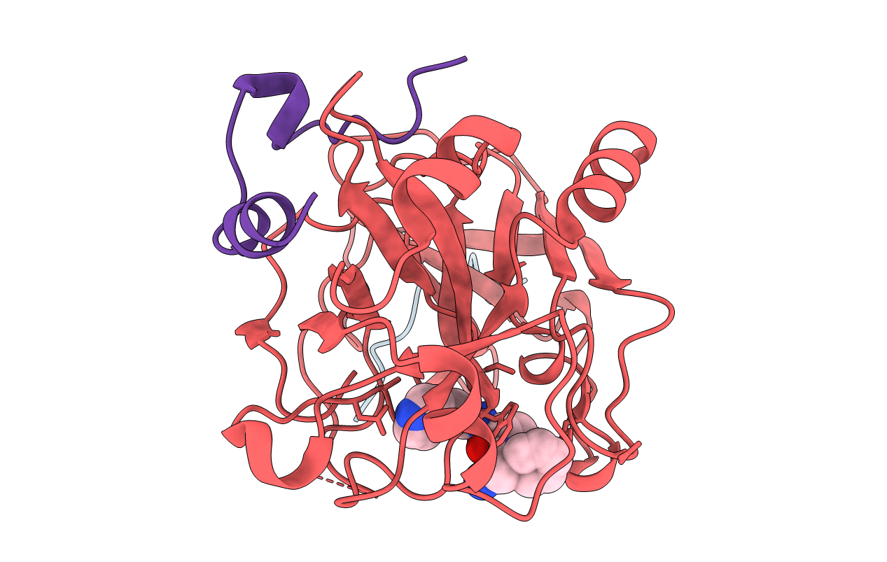
CRYSTAL STRUCTURE OF THROMBIN COMPLEXED WITH AN GUANIDINE-MIMETIC INHIBITOR
Biological Source:
Source Organism:
Homo sapiens (Taxon ID: 9606)
Hirudo medicinalis (Taxon ID: 6421)
Method Details:
Experimental Method:
Resolution:
1.80 Å
R-Value Work:
0.19
R-Value Observed:
0.19
Space Group:
C 1 2 1
Macromolecular Entities
Polymer Type:polypeptide(L)
Molecule:Hirudin
Chain IDs:C (auth: E)
Chain Length:11
Number of Molecules:1
Biological Source:Hirudo medicinalis
Polymer Type:polypeptide(L)
Molecule:THROMBIN (HEAVY CHAIN)
Gene (Uniprot):F2
Chain IDs:B (auth: H)
Chain Length:259
Number of Molecules:1
Biological Source:Homo sapiens
Polymer Type:polypeptide(L)
Molecule:THROMBIN (LIGHT CHAIN)
Gene (Uniprot):F2
Chain IDs:A (auth: L)
Chain Length:36
Number of Molecules:1
Biological Source:Homo sapiens
Modified Residue
Compound ID Chain ID Parent Comp ID Details 2D Image
TYS C TYR O-SULFO-L-TYROSINE
Ligand Molecules
Primary Citation
Structural analysis of thrombin complexed with potent inhibitors incorporating a phenyl group as a peptide mimetic and aminopyridines as guanidine substitutes.
J.Med.Chem. 41 2068 2075 (1998)
PMID: 9622548 DOI: 10.1021/jm970796l

Abstact

The structure of the noncovalent complex of human alpha-thrombin with a nonpeptide inhibitor containing a central phenyl scaffold, N-[2-[5-methyl-3-(2-chlorophenylsulfonyloxy)phenoxy]ethyl]-N- methyl-4 -aminopyridine (1), has been determined to 2.20 A resolution. In addition, the thrombin-bound structures of two distinct amino acid-based inhibitors (3 and 4) containing different aminopyridine-derived guanidine mimetics have been determined. Each compound occupies the same region of the active site and projects an aminopyridine, a central hydrophobic group, and an aryl group, into the S1, S2, and aryl subsites on thrombin. Nonpeptide 1 forms only one direct intermolecular hydrogen bond to the thrombin active site and forms no hydrogen bonds to ordered molecules of solvent. Close contacts are observed between main-chain carbonyl groups on thrombin and the edges of the central phenyl and aminopyridine rings and the sulfonyl group of 1 such that atoms carrying opposite partial charges are juxtaposed. Aminopyridine groups in 3 and 4 also form close contacts with the edges of carbonyl groups on thrombin and are flexibly accommodated in the S1 subsite. Superposition of the bound conformations of 1 and D-Phe-Pro-amidobutylguanidine (2) revealed that the central phenyl scaffold of 1 substitutes for the peptide main chain of 2.

Legend

Protein

Chemical

Disease

Primary Citation of related structures