1Q8M image
Deposition Date 2003-08-21
Release Date 2003-12-09
Last Version Date 2022-12-21
Entry Detail
PDB ID:
1Q8M
Title:
Crystal structure of the human myeloid cell activating receptor TREM-1
Biological Source:
Source Organism:
Homo sapiens (Taxon ID: 9606)
Host Organism:
Method Details:
Experimental Method:
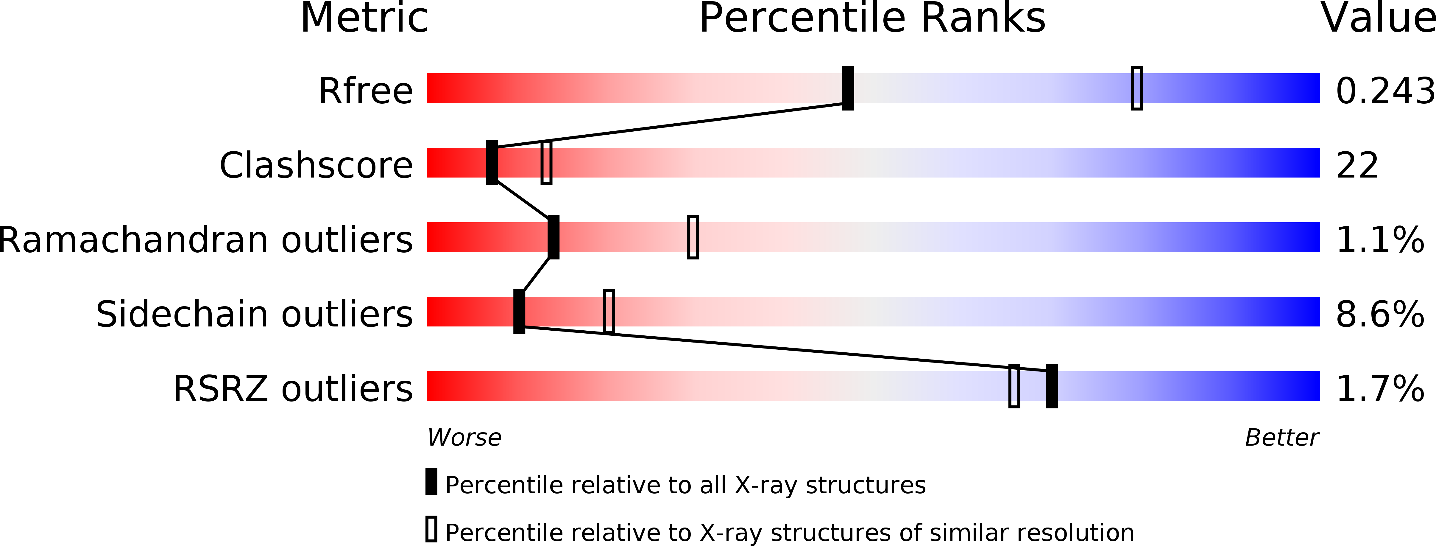
Resolution:
2.60 Å
R-Value Free:
0.25
R-Value Work:
0.20
Space Group:
P 1 21 1
Macromolecular Entities
Structures with similar UniProt ID
Protein Blast
Polymer Type:polypeptide(L)
Molecule:triggering receptor expressed on myeloid cells 1
Gene (Uniprot):TREM1
Chain IDs:A, B, C, D
Chain Length:127
Number of Molecules:4
Biological Source:Homo sapiens
Primary Citation
Crystal structure of the human myeloid cell activating receptor TREM-1
Structure 11 1527 1535 (2003)
PMID: 14656437 DOI: 10.1016/j.str.2003.11.001

Abstact

Triggering receptors expressed on myeloid cells (TREM) are a family of recently discovered receptors that play important roles in innate immune responses, such as to activate inflammatory responses and to contribute to septic shock in response to microbial-mediated infections. To date, two TREM receptors in human and several homologs in mice have been identified. We report the 2.6 A resolution crystal structure of the extracellular domain of human TREM-1. The overall fold of the receptor resembles that of a V-type immunoglobulin domain with differences primarily located in the N-terminal strand. TREM-1 forms a "head-to-tail" dimer with 4100 A(2) interface area that is partially mediated by a domain swapping between the first strands. This mode of dimer formation is different from the "head-to-head" dimerization that existed in V(H)V(L) domains of antibodies or V domains of T cell receptors. As a result, the dimeric TREM-1 most likely contains two distinct ligand binding sites.

Legend

Protein

Chemical

Disease

Primary Citation of related structures