1Q8F image
Deposition Date 2003-08-21
Release Date 2004-05-11
Last Version Date 2023-08-16
Entry Detail
PDB ID:
1Q8F
Keywords:
Title:
Crystal Structure of the E.coli pyrimidine nucleoside hydrolase yeiK
Biological Source:
Source Organism:
Escherichia coli (Taxon ID: 562)
Method Details:
Experimental Method:
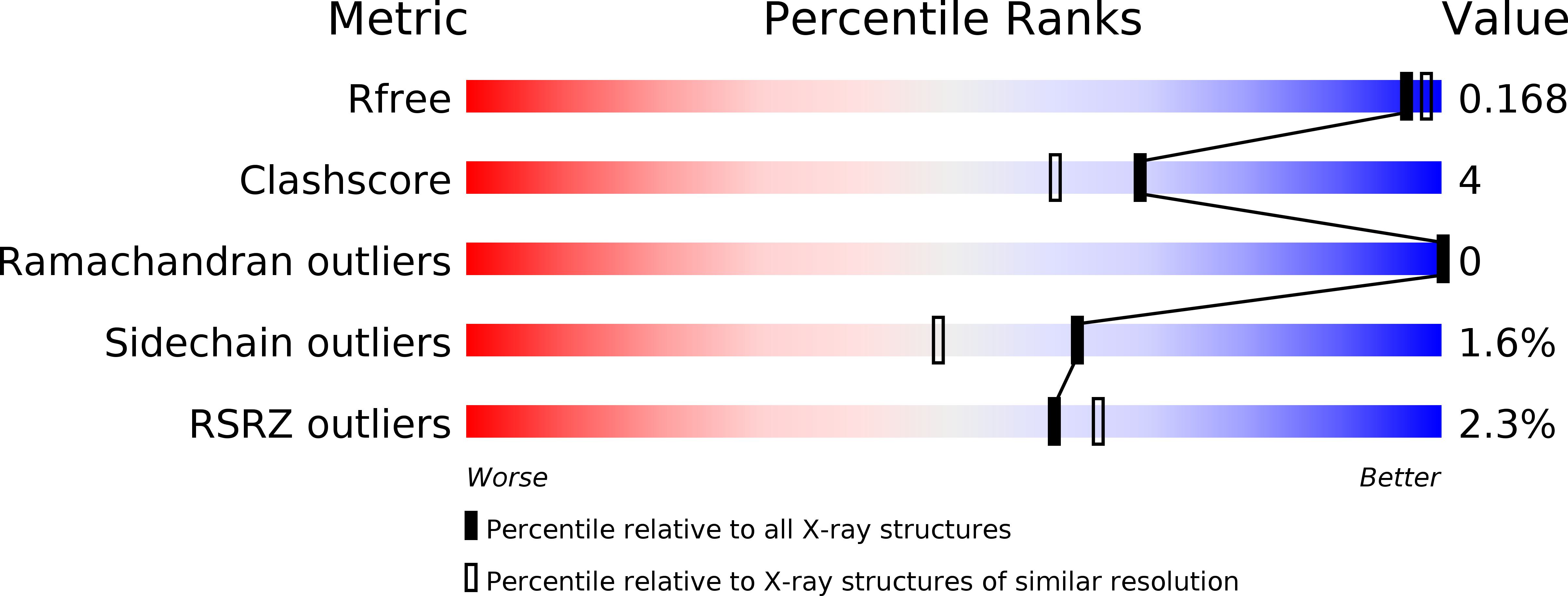
Resolution:
1.70 Å
R-Value Free:
0.16
R-Value Work:
0.15
R-Value Observed:
0.15
Space Group:
P 1
Macromolecular Entities
Polymer Type:polypeptide(L)
Molecule:Pyrimidine nucleoside hydrolase
Gene (Uniprot):rihB
Chain IDs:A, B, C, D
Chain Length:313
Number of Molecules:4
Biological Source:Escherichia coli
Primary Citation
Crystal structure to 1.7 a of the Escherichia coli pyrimidine nucleoside hydrolase YeiK, a novel candidate for cancer gene therapy.
STRUCTURE 12 739 749 (2004)
PMID: 15130467 DOI: 10.1016/j.str.2004.03.018

Abstact

Enzymes with nucleoside hydrolase (NH) activity are crucial for salvaging nucleic acid components in purine auxotrophic protozoan parasites, but are also present in prokaryotes and higher eukaryotes. Here we analyze the distribution of genes encoding for putative NH proteins and characterize the yeiK gene product from Escherichia coli as a pyrimidine-specific NH. The crystal structure of YeiK to 1.7 A defines the structural basis for its substrate specificity and identifies residues involved in the catalytic mechanism that differ from both nonspecific and purine-specific NHs. Large differences in the tetrameric quaternary structure compared to nonspecific protozoan NHs are brought forth by minor differences in the interacting surfaces. The first structural and functional characterization of a nonparasitic, pyrimidine nucleoside-specific NH suggests a possible role for these enzymes in the metabolism of tRNA nucleosides. The high catalytic efficiency of YeiK toward 5-fluorouridine could be exploited for suicide gene therapy in cancer treatment.

Legend

Protein

Chemical

Disease

Primary Citation of related structures