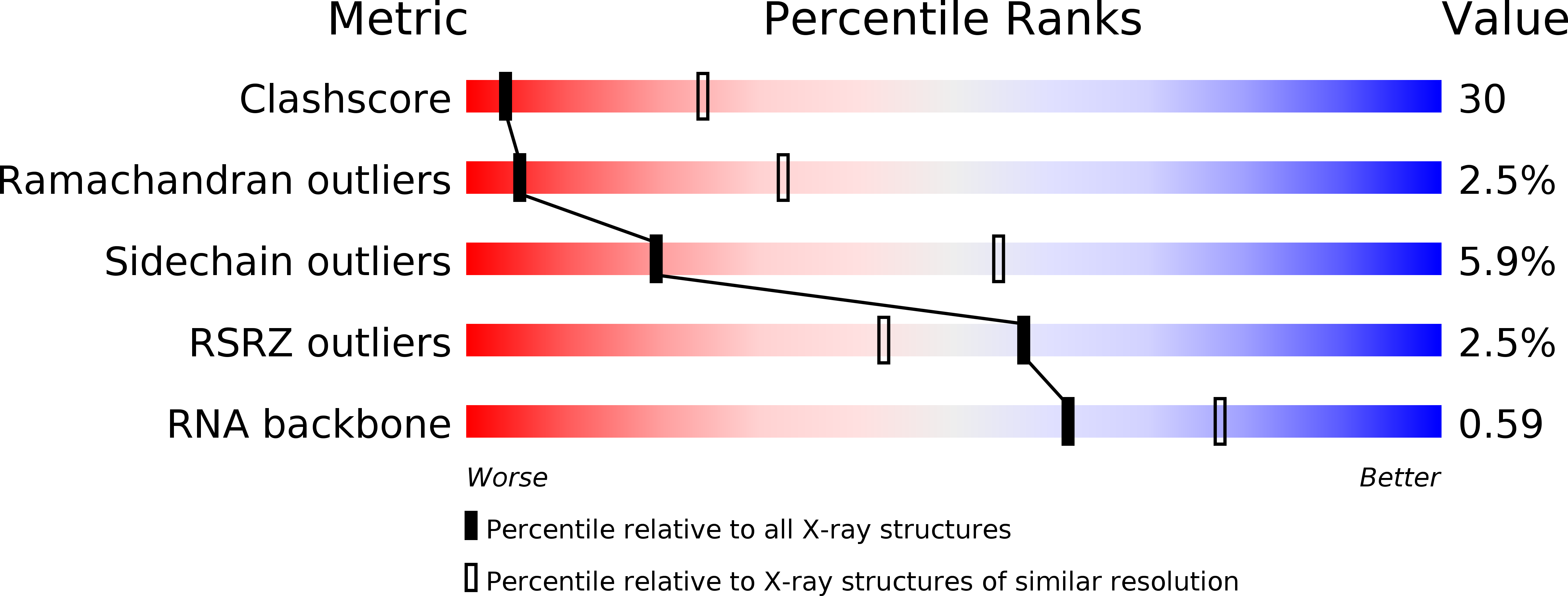
1Q7Y image
Entry Detail
PDB ID:
1Q7Y
Keywords:
Title:
Crystal Structure of CCdAP-Puromycin bound at the Peptidyl transferase center of the 50S ribosomal subunit
Biological Source:
Source Organism:
PDB Version:
Deposition Date:
2003-08-20
Release Date:
2003-10-07
Method Details:
Experimental Method:
Resolution:
3.20 Å
R-Value Free:
0.28
R-Value Work:
0.22
R-Value Observed:
0.22
Space Group:
C 2 2 21
Macromolecular Entities
Polymer Type:polypeptide(L)
Description:L37Ae 50S ribosomal protein
Chain IDs:BA (auth: 1)
Chain Length:73
Number of Molecules:1
Biological Source:Haloarcula marismortui
Polymer Type:polypeptide(L)
Description:50S ribosomal protein L37e
Chain IDs:CA (auth: 2)
Chain Length:56
Number of Molecules:1
Biological Source:Haloarcula marismortui
Polymer Type:polypeptide(L)
Description:50S ribosomal protein L39e
Chain IDs:DA (auth: 3)
Chain Length:48
Number of Molecules:1
Biological Source:Haloarcula marismortui
Polymer Type:polypeptide(L)
Description:50S ribosomal protein L44E
Chain IDs:EA (auth: 4)
Chain Length:92
Number of Molecules:1
Biological Source:Haloarcula marismortui
Polymer Type:polydeoxyribonucleotide/polyribonucleotide hybrid
Description:CCdA-P-Puromycin
Chain IDs:C (auth: 5)
Chain Length:3
Number of Molecules:1
Biological Source:
Polymer Type:polyribonucleotide
Description:23S ribosomal rna
Chain IDs:A
Chain Length:2922
Number of Molecules:1
Biological Source:Haloarcula marismortui
Polymer Type:polyribonucleotide
Description:5S ribosomal RNA
Chain IDs:B
Chain Length:122
Number of Molecules:1
Biological Source:Haloarcula marismortui
Polymer Type:polypeptide(L)
Description:50S ribosomal protein L2P
Chain IDs:D (auth: C)
Chain Length:239
Number of Molecules:1
Biological Source:Haloarcula marismortui
Polymer Type:polypeptide(L)
Description:50S ribosomal protein L3P
Chain IDs:E (auth: D)
Chain Length:337
Number of Molecules:1
Biological Source:Haloarcula marismortui
Polymer Type:polypeptide(L)
Description:50S ribosomal protein L4E
Chain IDs:F (auth: E)
Chain Length:246
Number of Molecules:1
Biological Source:Haloarcula marismortui
Polymer Type:polypeptide(L)
Description:50S ribosomal protein L5P
Chain IDs:G (auth: F)
Chain Length:176
Number of Molecules:1
Biological Source:Haloarcula marismortui
Polymer Type:polypeptide(L)
Description:50S ribosomal protein L6P
Chain IDs:H (auth: G)
Chain Length:177
Number of Molecules:1
Biological Source:Haloarcula marismortui
Polymer Type:polypeptide(L)
Description:50S ribosomal protein L7Ae
Chain IDs:I (auth: H)
Chain Length:119
Number of Molecules:1
Biological Source:Haloarcula marismortui
Polymer Type:polypeptide(L)
Description:Acidic ribosomal protein P0 homolog
Chain IDs:J (auth: I)
Chain Length:348
Number of Molecules:1
Biological Source:Haloarcula marismortui
Polymer Type:polypeptide(L)
Description:L10 Ribosomal Protein
Chain IDs:K (auth: J)
Chain Length:167
Number of Molecules:1
Biological Source:Haloarcula marismortui
Polymer Type:polypeptide(L)
Description:50S ribosomal protein L13P
Chain IDs:L (auth: K)
Chain Length:145
Number of Molecules:1
Biological Source:Haloarcula marismortui
Polymer Type:polypeptide(L)
Description:50S ribosomal protein L14P
Chain IDs:M (auth: L)
Chain Length:132
Number of Molecules:1
Biological Source:Haloarcula marismortui
Polymer Type:polypeptide(L)
Description:50S ribosomal protein L15P
Chain IDs:N (auth: M)
Chain Length:164
Number of Molecules:1
Biological Source:Haloarcula marismortui
Polymer Type:polypeptide(L)
Description:L15 Ribosomal Protein
Chain IDs:O (auth: N)
Chain Length:194
Number of Molecules:1
Biological Source:Haloarcula marismortui
Polymer Type:polypeptide(L)
Description:50S ribosomal protein L18P
Chain IDs:P (auth: O)
Chain Length:186
Number of Molecules:1
Biological Source:Haloarcula marismortui
Polymer Type:polypeptide(L)
Description:50S ribosomal protein L18e
Chain IDs:Q (auth: P)
Chain Length:115
Number of Molecules:1
Biological Source:Haloarcula marismortui
Polymer Type:polypeptide(L)
Description:50S ribosomal protein L19E
Chain IDs:R (auth: Q)
Chain Length:148
Number of Molecules:1
Biological Source:Haloarcula marismortui
Polymer Type:polypeptide(L)
Description:50S ribosomal protein L21e
Chain IDs:S (auth: R)
Chain Length:95
Number of Molecules:1
Biological Source:Haloarcula marismortui
Polymer Type:polypeptide(L)
Description:50S ribosomal protein L22P
Chain IDs:T (auth: S)
Chain Length:154
Number of Molecules:1
Biological Source:Haloarcula marismortui
Polymer Type:polypeptide(L)
Description:50S ribosomal protein L23P
Chain IDs:U (auth: T)
Chain Length:84
Number of Molecules:1
Biological Source:Haloarcula marismortui
Polymer Type:polypeptide(L)
Description:50S ribosomal protein L24P
Chain IDs:V (auth: U)
Chain Length:119
Number of Molecules:1
Biological Source:Haloarcula marismortui
Polymer Type:polypeptide(L)
Description:50S ribosomal protein L24E
Chain IDs:W (auth: V)
Chain Length:66
Number of Molecules:1
Biological Source:Haloarcula marismortui
Polymer Type:polypeptide(L)
Description:50S ribosomal protein L29P
Chain IDs:X (auth: W)
Chain Length:70
Number of Molecules:1
Biological Source:Haloarcula marismortui
Polymer Type:polypeptide(L)
Description:50S ribosomal protein L30P
Chain IDs:Y (auth: X)
Chain Length:154
Number of Molecules:1
Biological Source:Haloarcula marismortui
Polymer Type:polypeptide(L)
Description:50S ribosomal protein L31e
Chain IDs:Z (auth: Y)
Chain Length:91
Number of Molecules:1
Biological Source:Haloarcula marismortui
Polymer Type:polypeptide(L)
Description:50S ribosomal protein L32E
Chain IDs:AA (auth: Z)
Chain Length:240
Number of Molecules:1
Biological Source:Haloarcula marismortui
Primary Citation
Structural Insights Into Peptide Bond Formation
Proc.Natl.Acad.Sci.USA 99 11670 11675 (2002)
PMID: 12185246 DOI: 10.1073/pnas.172404099

Abstact

The large ribosomal subunit catalyzes peptide bond formation and will do so by using small aminoacyl- and peptidyl-RNA fragments of tRNA. We have refined at 3-A resolution the structures of both A and P site substrate and product analogues, as well as an intermediate analogue, bound to the Haloarcula marismortui 50S ribosomal subunit. A P site substrate, CCA-Phe-caproic acid-biotin, binds equally to both sites, but in the presence of sparsomycin binds only to the P site. The CCA portions of these analogues are bound identically by either the A or P loop of the 23S rRNA. Combining the separate P and A site substrate complexes into one model reveals interactions that may occur when both are present simultaneously. The alpha-NH(2) group of an aminoacylated fragment in the A site forms one hydrogen bond with the N3 of A2486 (2451) and may form a second hydrogen bond either with the 2' OH of the A-76 ribose in the P site or with the 2' OH of A2486 (2451). These interactions position the alpha amino group adjacent to the carbonyl carbon of esterified P site substrate in an orientation suitable for a nucleophilic attack.

Legend

Protein

Chemical

Disease

Primary Citation of related structures