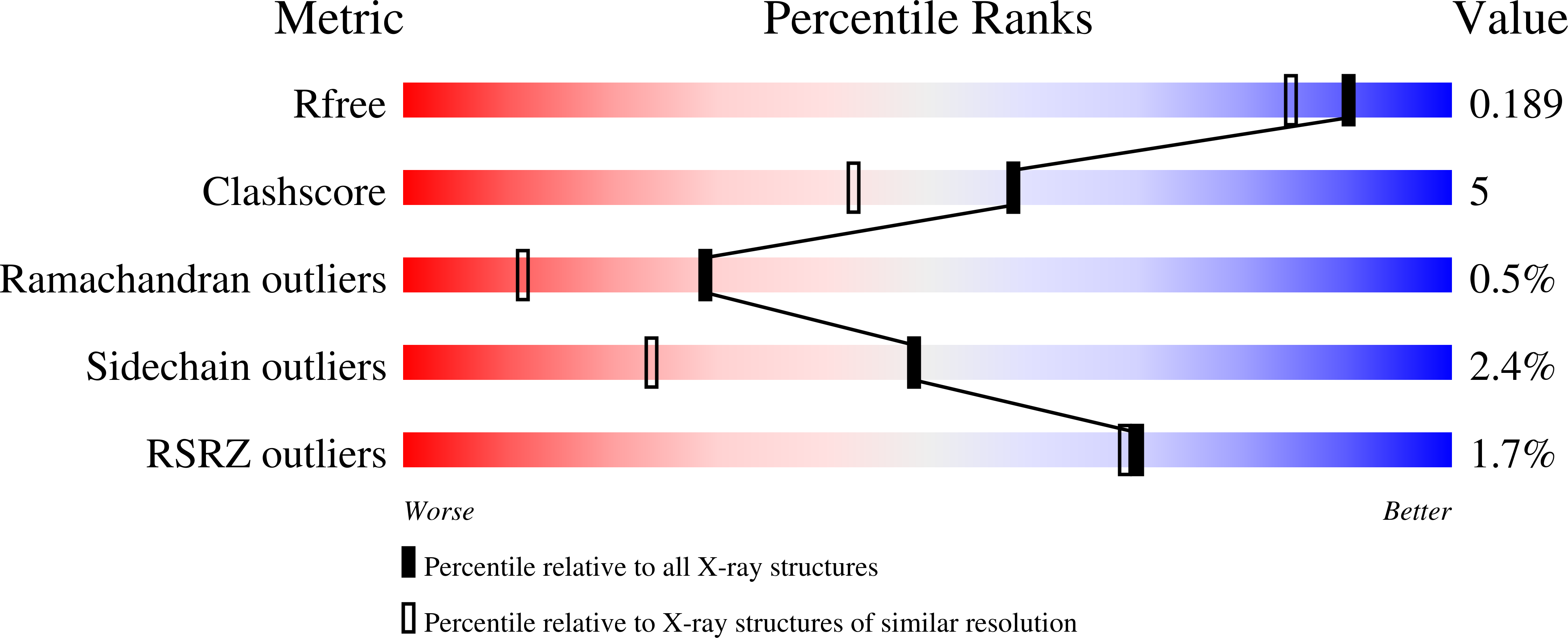
1Q7E image
Deposition Date 2003-08-18
Release Date 2003-09-30
Last Version Date 2024-11-20
Entry Detail
PDB ID:
1Q7E
Keywords:
Title:
Crystal Structure of YfdW protein from E. coli
Biological Source:
Source Organism:
Escherichia coli (Taxon ID: 562)
Host Organism:
Method Details:
Experimental Method:
Resolution:
1.60 Å
R-Value Free:
0.18
R-Value Work:
0.15
Space Group:
C 1 2 1
Macromolecular Entities
Structures with similar UniProt ID
Protein Blast
Polymer Type:polypeptide(L)
Molecule:Hypothetical protein yfdW
Gene (Uniprot):frc
Chain IDs:A
Chain Length:428
Number of Molecules:1
Biological Source:Escherichia coli
Modified Residue
Compound ID Chain ID Parent Comp ID Details 2D Image
MSE A MET SELENOMETHIONINE
Primary Citation
Structure of Escherichia coli YfdW, a type III CoA transferase.
Acta Crystallogr.,Sect.D 60 507 511 (2004)
PMID: 14993676 DOI: 10.1107/S0907444904000034

Abstact

Crystal structures are reported for free and coenzyme A (CoA) bound forms of the YfdW protein from Escherichia coli, a representative type III CoA transferase. The structures reveal a two-domain protomer with interdomain connections forming a ring-like structure with a large central hole. Two protomers associate to form a highly intertwined dimer in which the hole of each ring is filled by the partner molecule. Each protomer binds a single CoA molecule and these CoA-binding sites are distant from one another in the dimer.

Legend

Protein

Chemical

Disease

Primary Citation of related structures