1Q5Z image
Deposition Date 2003-08-11
Release Date 2003-10-07
Last Version Date 2024-02-14
Entry Detail
PDB ID:
1Q5Z
Keywords:
Title:
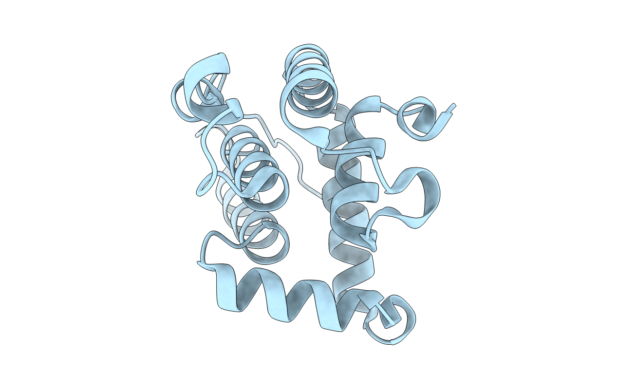
Crystal Structure of the C-terminal Actin Binding Domain of Salmonella Invasion Protein A (SipA)
Biological Source:
Source Organism:
Method Details:
Experimental Method:
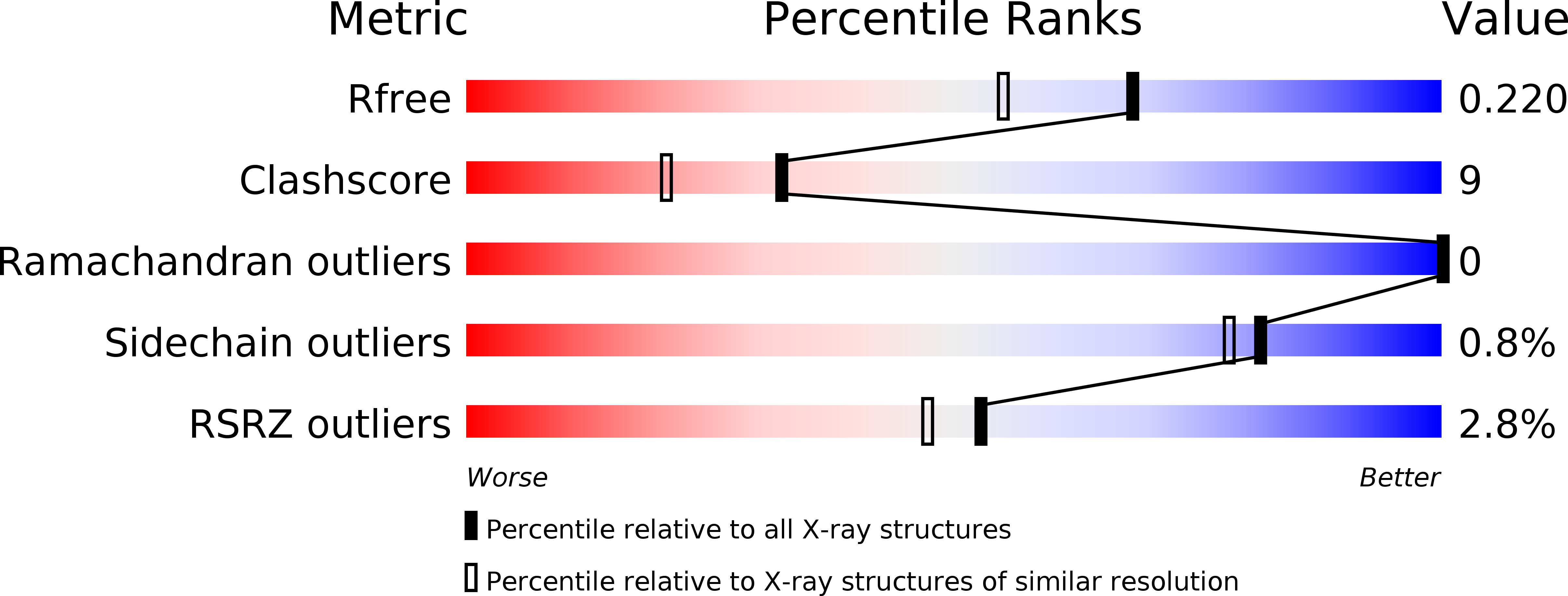
Resolution:
1.80 Å
R-Value Free:
0.22
R-Value Work:
0.19
R-Value Observed:
0.19
Space Group:
P 43 21 2
Macromolecular Entities
Structures with similar UniProt ID
Protein Blast
Polymer Type:polypeptide(L)
Molecule:SipA
Chain IDs:A
Chain Length:177
Number of Molecules:1
Biological Source:Salmonella typhimurium
Primary Citation
Salmonella SipA polymerizes actin by stapling filaments with nonglobular protein arms.
Science 301 1918 1921 (2003)
PMID: 14512630 DOI: 10.1126/science.1088433

Abstact

Like many bacterial pathogens, Salmonella spp. use a type III secretion system to inject virulence proteins into host cells. The Salmonella invasion protein A (SipA) binds host actin, enhances its polymerization near adherent extracellular bacteria, and contributes to cytoskeletal rearrangements that internalize the pathogen. By combining x-ray crystallography of SipA with electron microscopy and image analysis of SipA-actin filaments, we show that SipA functions as a "molecular staple," in which a globular domain and two nonglobular "arms" mechanically stabilize the filament by tethering actin subunits in opposing strands. Deletion analysis of the tethering arms provides strong support for this model.

Legend

Protein

Chemical

Disease

Primary Citation of related structures
Feedback Form
Name
Email
Institute
Feedback