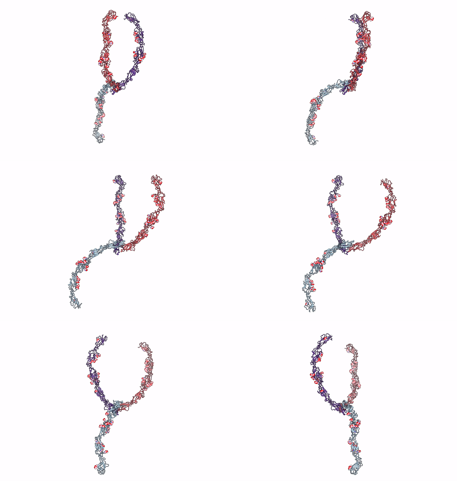
1Q5B image
Deposition Date 2003-08-06
Release Date 2003-10-07
Last Version Date 2024-12-25
Entry Detail
PDB ID:
1Q5B
Title:
lambda-shaped TRANS and CIS interactions of cadherins model based on fitting C-cadherin (1L3W) to 3D map of desmosomes obtained by electron tomography
Biological Source:
Source Organism:
Mus musculus (Taxon ID: 10090)
Method Details:
Experimental Method:
Resolution:
30.00 Å
Aggregation State:
TISSUE
Reconstruction Method:
TOMOGRAPHY
Macromolecular Entities
Polymer Type:polypeptide(L)
Molecule:EP-cadherin
Chain IDs:A, B, C
Chain Length:880
Number of Molecules:3
Biological Source:Mus musculus
Modified Residue
Compound ID Chain ID Parent Comp ID Details 2D Image
ASN A ASN GLYCOSYLATION SITE
THR A THR GLYCOSYLATION SITE
Primary Citation
Untangling Desmosomal Knots with Electron Tomography
Science 302 109 113 (2003)
PMID: 14526082 DOI: 10.1126/science.1086957

Abstact

Cell adhesion by adherens junctions and desmosomes relies on interactions between cadherin molecules. However, the molecular interfaces that define molecular specificity and that mediate adhesion remain controversial. We used electron tomography of plastic sections from neonatal mouse skin to visualize the organization of desmosomes in situ. The resulting three-dimensional maps reveal individual cadherin molecules forming discrete groups and interacting through their tips. Fitting of an x-ray crystal structure for C-cadherin to these maps is consistent with a flexible intermolecular interface mediated by an exchange of amino-terminal tryptophans. This flexibility suggests a novel mechanism for generating both cis and trans interactions and for propagating these adhesive interactions along the junction.

Legend

Protein

Chemical

Disease

Primary Citation of related structures
Feedback Form
Name
Email
Institute
Feedback