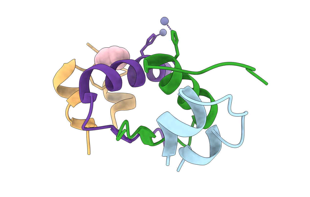
1Q4V image
Deposition Date 2003-08-04
Release Date 2003-08-19
Last Version Date 2024-11-20
Entry Detail
PDB ID:
1Q4V
Title:
CRYSTAL STRUCTURE OF ALLO-ILEA2-INSULIN, AN INACTIVE CHIRAL ANALOGUE: IMPLICATIONS FOR THE MECHANISM OF RECEPTOR
Biological Source:
Source Organism:
(Taxon ID: )
Method Details:
Experimental Method:
Resolution:
2.00 Å
R-Value Free:
0.26
R-Value Work:
0.21
Space Group:
H 3
Macromolecular Entities
Polymer Type:polypeptide(L)
Molecule:Insulin
Chain IDs:A, C
Chain Length:21
Number of Molecules:2
Biological Source:
Polymer Type:polypeptide(L)
Molecule:Insulin
Chain IDs:B, D
Chain Length:30
Number of Molecules:2
Biological Source:
Modified Residue
Compound ID Chain ID Parent Comp ID Details 2D Image
IIL A ILE ISO-ISOLEUCINE
Primary Citation
Crystal structure of allo-Ile(A2)-insulin, an inactive chiral analogue: implications for the mechanism of receptor binding.
Biochemistry 42 12770 12783 (2003)
PMID: 14596591 DOI: 10.1021/bi034430o

Abstact

The crystal structure of an inactive chiral analogue of insulin containing nonstandard substitution allo-Ile(A2) is described at 2.0 A resolution. In native insulin, the invariant Ile(A2) side chain anchors the N-terminal alpha-helix of the A-chain to the hydrophobic core. The structure of the variant protein was determined by molecular replacement as a T(3)R(3) zinc hexamer. Whereas respective T- and R-state main-chain structures are similar to those of native insulin (main-chain root-mean-square deviations (RMSD) of 0.45 and 0.54 A, respectively), differences in core packing are observed near the variant side chain. The R-state core resembles that of the native R-state with a local inversion of A2 orientation (core side chain RMSD 0.75 A excluding A2); in the T-state, allo-Ile(A2) exhibits an altered conformation in association with the reorganization of the surrounding side chains (RMSD 0.98 A). Surprisingly, the core of the R-state is similar to that observed in solution nuclear magnetic resonance (NMR) studies of an engineered T-like monomer containing the same chiral substitution (allo-Ile(A2)-DKP-insulin; Xu, B., Hua, Q. X., Nakagawa, S. H., Jia, W., Chu, Y. C., Katsoyannis, P. G., and Weiss, M. A. (2002) J. Mol. Biol. 316, 435-441). Simulation of NOESY spectra based on crystallographic protomers enables the analysis of similarities and differences in solution. The different responses of the T- and R-state cores to chiral perturbation illustrates both their intrinsic plasticity and constraints imposed by hexamer assembly. Although variant T- and R-protomers retain nativelike protein surfaces, the receptor-binding activity of allo-Ile(A2)-insulin is low (2% relative to native insulin). This seeming paradox suggests that insulin undergoes a change in conformation to expose Ile(A2) at the hormone-receptor interface.

Legend

Protein

Chemical

Disease

Primary Citation of related structures