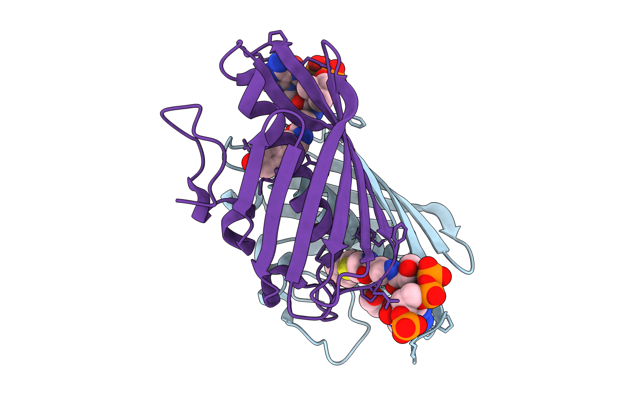
1Q4U image
Deposition Date 2003-08-04
Release Date 2003-09-23
Last Version Date 2023-08-16
Entry Detail
PDB ID:
1Q4U
Keywords:
Title:
Crystal structure of 4-hydroxybenzoyl CoA thioesterase from arthrobacter sp. strain SU complexed with 4-hydroxybenzyl CoA
Biological Source:
Source Organism:
Host Organism:
Method Details:
Experimental Method:
Resolution:
1.60 Å
R-Value Free:
0.21
R-Value Work:
0.17
R-Value Observed:
0.17
Space Group:
P 32 2 1
Macromolecular Entities
Polymer Type:polypeptide(L)
Molecule:Thioesterase
Gene (Uniprot):fcbC
Chain IDs:A, B
Chain Length:151
Number of Molecules:2
Biological Source:Arthrobacter sp.
Primary Citation
The Structure of 4-Hydroxybenzoyl-CoA Thioesterase from Arthrobacter sp. strain SU
J.Biol.Chem. 278 43709 43716 (2003)
PMID: 12907670 DOI: 10.1074/jbc.M308198200

Abstact

The 4-chlorobenzoyl-CoA dehalogenation pathway in certain Arthrobacter and Pseudomonas bacterial species contains three enzymes: a ligase, a dehalogenase, and a thioesterase. Here we describe the high resolution x-ray crystallographic structure of the 4-hydroxybenzoyl-CoA thioesterase from Arthrobacter sp. strain SU. The tetrameric enzyme is a dimer of dimers with each subunit adopting the so-called "hot dog fold" composed of six strands of anti-parallel beta-sheet flanked on one side by a rather long alpha-helix. The dimers come together to form the tetramer with their alpha-helices facing outwards. This quaternary structure is in sharp contrast to that previously observed for the 4-hydroxybenzoyl-CoA thioesterase from Pseudomonas species strain CBS-3, whereby the dimers forming the tetramer pack with their alpha-helices projecting toward the interfacial region. In the Arthrobacter thioesterase, each of the four active sites is formed by three of the subunits of the tetramer. On the basis of both structural and kinetic data, it appears that Glu73 is the active site base in the Arthrobacter thioesterase. Remarkably, this residue is located on the opposite side of the substrate-binding pocket compared with that observed for the Pseudomonas enzyme. Although these two bacterial thioesterases demonstrate equivalent catalytic efficiencies, substrate specificities, and metabolic functions, their quaternary structures, CoA-binding sites, and catalytic platforms are decidedly different.

Legend

Protein

Chemical

Disease

Primary Citation of related structures