Deposition Date
2003-08-01
Release Date
2004-08-03
Last Version Date
2024-11-06
Entry Detail
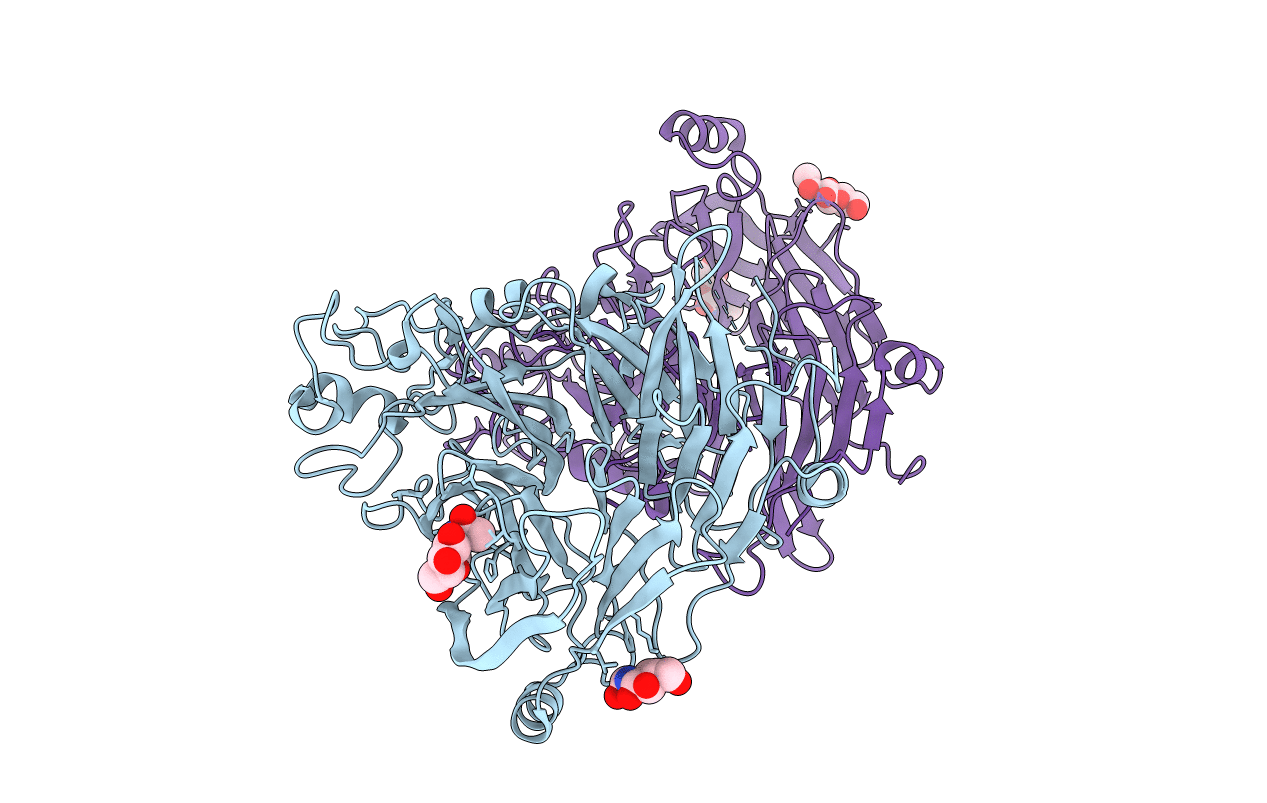
PDB ID:
1Q47
Keywords:
Title:
Structure of the Semaphorin 3A Receptor-Binding Module
Biological Source:
Source Organism(s):
Mus musculus (Taxon ID: 10090)
Expression System(s):
Method Details:
Experimental Method:
Resolution:
2.80 Å
R-Value Free:
0.29
R-Value Observed:
0.24
Space Group:
C 1 2 1