1Q46 image
Deposition Date 2003-08-01
Release Date 2003-10-28
Last Version Date 2024-04-03
Entry Detail
PDB ID:
1Q46
Keywords:
Title:
crystal structure of the eIF2 alpha subunit from saccharomyces cerevisia
Biological Source:
Source Organism:
Method Details:
Experimental Method:
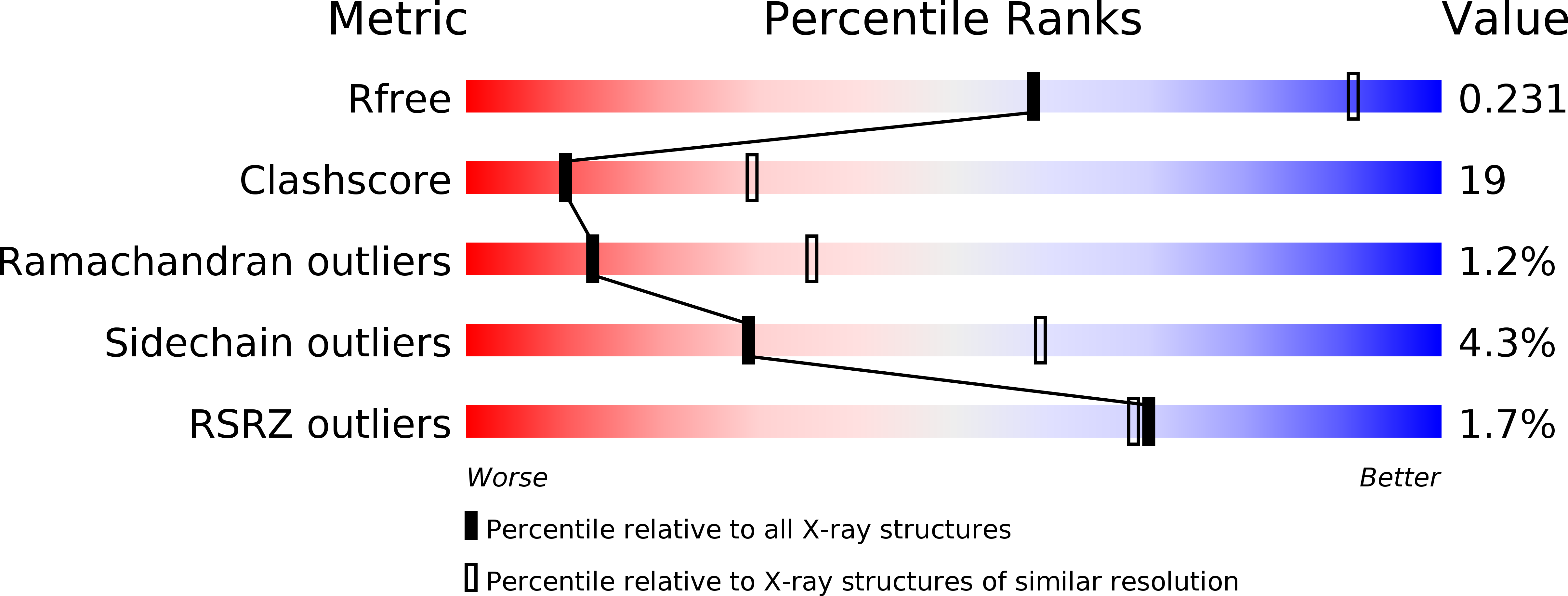
Resolution:
2.86 Å
R-Value Free:
0.23
R-Value Work:
0.21
R-Value Observed:
0.22
Space Group:
P 43 21 2
Macromolecular Entities
Structures with similar UniProt ID
Protein Blast
Polymer Type:polypeptide(L)
Molecule:translation initiation factor 2 alpha subunit
Gene (Uniprot):SUI2
Chain IDs:A
Chain Length:175
Number of Molecules:1
Biological Source:Saccharomyces cerevisiae
Ligand Molecules
Primary Citation
The crystal structure of the N-terminal region of the alpha subunit of translation initiation factor 2 (eIF2alpha) from Saccharomyces cerevisiae provides a view of the loop containing serine 51, the target of the eIF2alpha-specific kinases.
J.Mol.Biol. 334 187 195 (2003)
PMID: 14607111 DOI: 10.1016/j.jmb.2003.09.045

Abstact

The alpha subunit of translation initiation factor 2 (eIF2alpha) is the target of specific kinases that can phosphorylate a conserved serine residue as part of a mechanism for regulating protein expression at the translational level in eukaryotes. The structure of the 20 kDa N-terminal region of eIF2alpha from Saccharomyces cerevisiae was determined by X-ray crystallography at 2.5A resolution. In most respects, the structure is similar to that of the recently solved human eIF2alpha; the rather elongated protein contains a five-stranded antiparallel beta-barrel in its N-terminal region, followed by an almost entirely helical domain. The S.cerevisiae eIF2alpha lacks a disulfide bridge that is present in the homologous protein in humans and some of the other higher eukaryotes. Interestingly, a conserved loop consisting of residues 51-65 and containing serine 51, the putative phosphorylation site, is visible in the electron density maps of the S.cerevisiae eIF2alpha; most of this functionally important loop was not observed in the crystal structure of the human protein. This loop is relatively exposed to solvent, and contains two short 3(10) helices in addition to some extended structure. Serine 51 is located at the C-terminal end of one of the 3(10) helices and near several conserved positively charged residues. The side-chain of serine 51 is sufficiently exposed so that its phosphorylation would not necessitate a substantial change in the protein structure. The structures and relative positions of residues that have been implicated in kinase binding and in the interaction with guanine nucleotide exchange factor (eIF2B) are described.

Legend

Protein

Chemical

Disease

Primary Citation of related structures