1Q3M image
Deposition Date 2003-07-30
Release Date 2003-09-16
Last Version Date 2022-03-02
Entry Detail
PDB ID:
1Q3M
Title:
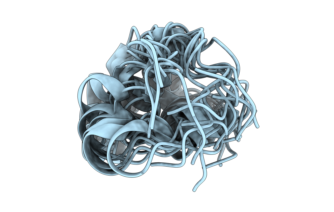
1H NMR structure bundle of bovine Ca2+-osteocalcin
Biological Source:
Source Organism:
Bos taurus (Taxon ID: 9913)
Method Details:
Experimental Method:
Conformers Submitted:
13
Selection Criteria:
structures with the lowest energy
Macromolecular Entities
Structures with similar UniProt ID
Protein Blast
Polymer Type:polypeptide(L)
Molecule:Osteocalcin
Gene (Uniprot):BGLAP
Chain IDs:A
Chain Length:49
Number of Molecules:1
Biological Source:Bos taurus
Modified Residue
Compound ID Chain ID Parent Comp ID Details 2D Image
CGU A GLU GAMMA-CARBOXY-GLUTAMIC ACID
Ligand Molecules
Primary Citation
The Three-Dimensional Structure of Bovine Calcium Ion-Bound Osteocalcin Using 1H NMR Spectroscopy
Biochemistry 42 7769 7779 (2003)
PMID: 12820886 DOI: 10.1021/bi034470s

Abstact

Structural information on osteocalcin or other noncollagenous bone proteins is very limited. We have solved the three-dimensional structure of calcium bound osteocalcin using (1)H 2D NMR techniques and proposed a mechanism for mineral binding. The protons in the 49 amino acid sequence were assigned using standard two-dimensional homonuclear NMR experiments. Distance constraints, dihedral angle constraints, hydrogen bonds, and (1)H and (13)C chemical shifts were all used to calculate a family of 13 structures. The tertiary structure of the protein consisted of an unstructured N terminus and a C-terminal loop (residues 16-49) formed by long-range hydrophobic interactions. Elements of secondary structure within residues 16-49 include type III turns (residues 20-25) and two alpha-helical regions (residues 27-35 and 41-44). The three Gla residues project from the same face of the helical turns and are surface exposed. The genetic algorithm-molecular dynamics simulation approach was used to place three calcium atoms on the NMR-derived structure. One calcium atom was coordinated by three side chain oxygen atoms, two from Asp30, and one from Gla24. The second calcium atom was coordinated to four oxygen atoms, two from the side chain in Gla 24, and two from the side chain of Gla 21. The third calcium atom was coordinated to two oxygen atoms of the side chain of Gla17. The best correlation of the distances between the uncoordinated Gla oxygen atoms is with the intercalcium distance of 9.43 A in hydroxyapatite. The structure may provide further insight into the function of osteocalcin.

Legend

Protein

Chemical

Disease

Primary Citation of related structures
Feedback Form
Name
Email
Institute
Feedback