1Q3F image
Deposition Date 2003-07-29
Release Date 2004-03-23
Last Version Date 2023-08-16
Entry Detail
PDB ID:
1Q3F
Keywords:
Title:
Uracil DNA glycosylase bound to a cationic 1-aza-2'-deoxyribose-containing DNA
Biological Source:
Source Organism:
Homo sapiens (Taxon ID: 9606)
Host Organism:
Method Details:
Experimental Method:
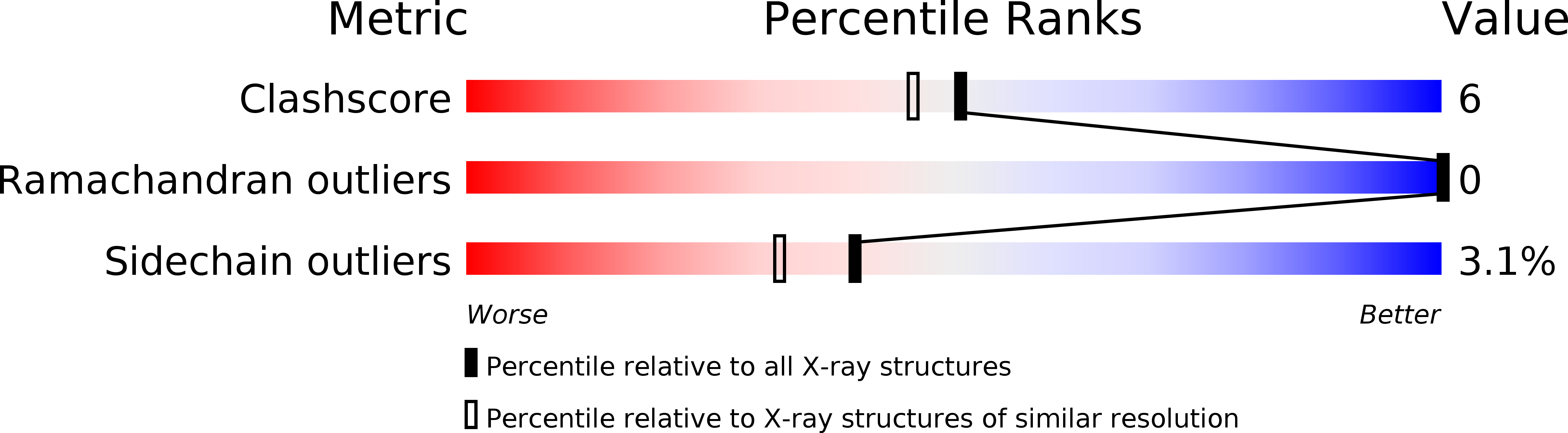
Resolution:
1.90 Å
R-Value Free:
0.23
R-Value Work:
0.19
R-Value Observed:
0.19
Space Group:
P 21 21 21
Macromolecular Entities
Polymer Type:polypeptide(L)
Molecule:Uracil-DNA glycosylase
Gene (Uniprot):UNG
Chain IDs:C (auth: A)
Chain Length:223
Number of Molecules:1
Biological Source:Homo sapiens
Primary Citation
Electrostatic guidance of glycosyl cation migration along the reaction coordinate of uracil DNA glycosylase.
Biochemistry 42 12455 12460 (2003)
PMID: 14580190 DOI: 10.1021/bi035372+

Abstact

The DNA repair enzyme uracil DNA glycosylase has been crystallized with a cationic 1-aza-2'-deoxyribose-containing DNA that mimics the ultimate transition state of the reaction in which the water nucleophile attacks the anomeric center of the oxacarbenium ion-uracil anion reaction intermediate. Comparison with substrate and product structures, and the previous structure of the intermediate determined by kinetic isotope effects, reveals an exquisite example of geometric strain, least atomic motion, and electrophile migration in biological catalysis. This structure provides a rare opportunity to reconstruct the detailed structural transformations that occur along an enzymatic reaction coordinate.

Legend

Protein

Chemical

Disease

Primary Citation of related structures