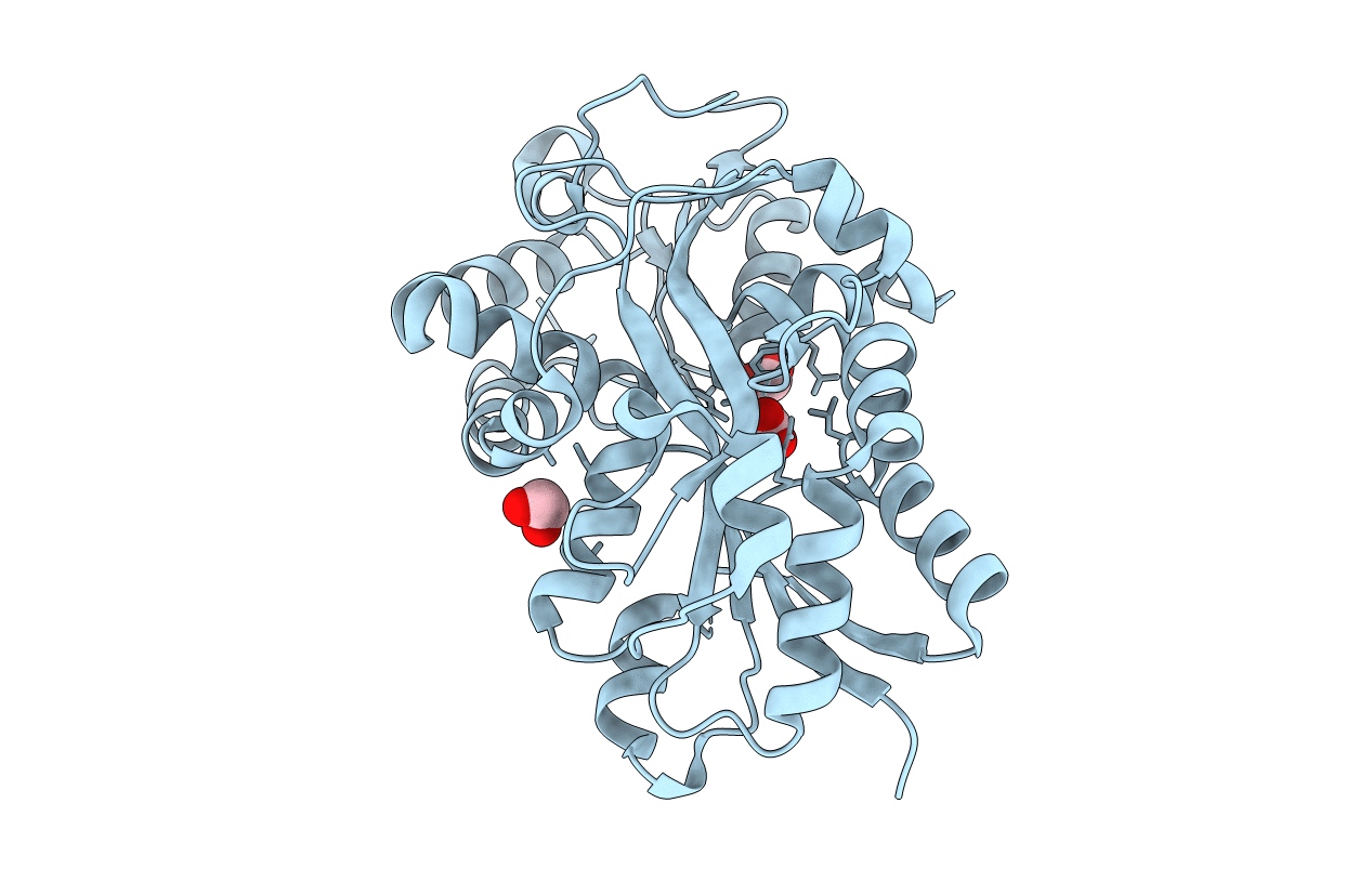
1Q35 image
Entry Detail
PDB ID:
1Q35
Title:
Crystal Structure of Pasteurella haemolytica Apo Ferric ion-Binding Protein A
Biological Source:
Source Organism:
Host Organism:
PDB Version:
Deposition Date:
2003-07-28
Release Date:
2003-11-11
Method Details:
Experimental Method:
Resolution:
1.20 Å
R-Value Free:
0.19
R-Value Work:
0.17
R-Value Observed:
0.17
Space Group:
C 2 2 21
Macromolecular Entities
Polymer Type:polypeptide(L)
Description:iron binding protein FbpA
Chain IDs:A
Chain Length:320
Number of Molecules:1
Biological Source:Mannheimia haemolytica
Primary Citation
Crystal structure of Pasteurella haemolytica ferric ion-binding protein A reveals a novel class of bacterial iron-binding proteins
J.Biol.Chem. 278 41093 41098 (2003)
PMID: 12882966 DOI: 10.1074/jbc.M306821200

Abstact

Pasteurellosis caused by the Gram-negative pathogen Pasteurella haemolytica is a serious disease leading to death in cattle. To scavenge growth-limiting iron from the host, the pathogen utilizes the periplasmic ferric ion-binding protein A (PhFbpA) as a component of an ATP-binding cassette transport pathway. We report the 1.2-A structure of the iron-free (apo) form of PhFbpA, which is a member of the transferrin structural superfamily. The protein structure adopts a closed conformation, allowing us to reliably assign putative iron-coordinating residues. Based on our analysis, PhFbpA utilizes a unique constellation of binding site residues and anions to octahedrally coordinate an iron atom. A surprising finding in the structure is the presence of two formate anions on opposite sides of the iron-binding pocket. The formate ions tether the N- and C-terminal domains of the protein and stabilize the closed structure, also providing clues as to probable candidates for synergistic anions in the iron-loaded state. PhFbpA represents a new class of bacterial iron-binding proteins.

Legend

Protein

Chemical

Disease

Primary Citation of related structures