1Q31 image
Entry Detail
PDB ID:
1Q31
Title:
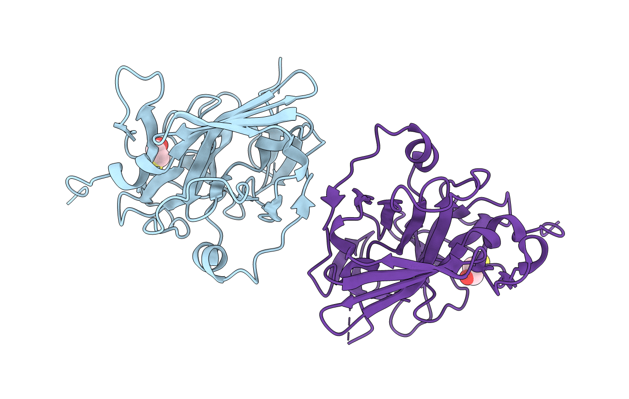
Crystal Structure of the Tobacco Etch Virus Protease C151A mutant
Biological Source:
Source Organism:
Host Organism:
PDB Version:
Deposition Date:
2003-07-28
Release Date:
2004-11-02
Method Details:
Experimental Method:
Resolution:
2.70 Å
R-Value Free:
0.30
R-Value Work:
0.24
R-Value Observed:
0.24
Space Group:
C 1 2 1
Macromolecular Entities
Polymer Type:polypeptide(L)
Description:Nuclear inclusion protein A
Mutations:C151A
Chain IDs:A, B
Chain Length:242
Number of Molecules:2
Biological Source:Tobacco etch virus
Ligand Molecules
Primary Citation
Crystal structure of tobacco etch virus protease shows the protein C terminus bound within the active site.
J.Mol.Biol. 350 145 155 (2005)
PMID: 15919091 DOI: 10.1016/j.jmb.2005.04.013

Abstact

Tobacco etch virus (TEV) protease is a cysteine protease exhibiting stringent sequence specificity. The enzyme is widely used in biotechnology for the removal of the affinity tags from recombinant fusion proteins. Crystal structures of two TEV protease mutants as complexes with a substrate and a product peptide provided the first insight into the mechanism of substrate specificity of this enzyme. We now report a 2.7A crystal structure of a full-length inactive C151A mutant protein crystallised in the absence of peptide. The structure reveals the C terminus of the protease bound to the active site. In addition, we determined dissociation constants of TEV protease substrate and product peptides using isothermal titration calorimetry for various forms of this enzyme. Data suggest that TEV protease could be inhibited by the peptide product of autolysis. Separate modes of recognition for native substrates and the site of TEV protease self-cleavage are proposed.

Legend

Protein

Chemical

Disease

Primary Citation of related structures