1Q2V image
Deposition Date 2003-07-26
Release Date 2004-01-27
Last Version Date 2024-02-14
Entry Detail
PDB ID:
1Q2V
Keywords:
Title:
Crystal structure of the chaperonin from Thermococcus strain KS-1 (nucleotide-free form)
Biological Source:
Source Organism:
Method Details:
Experimental Method:
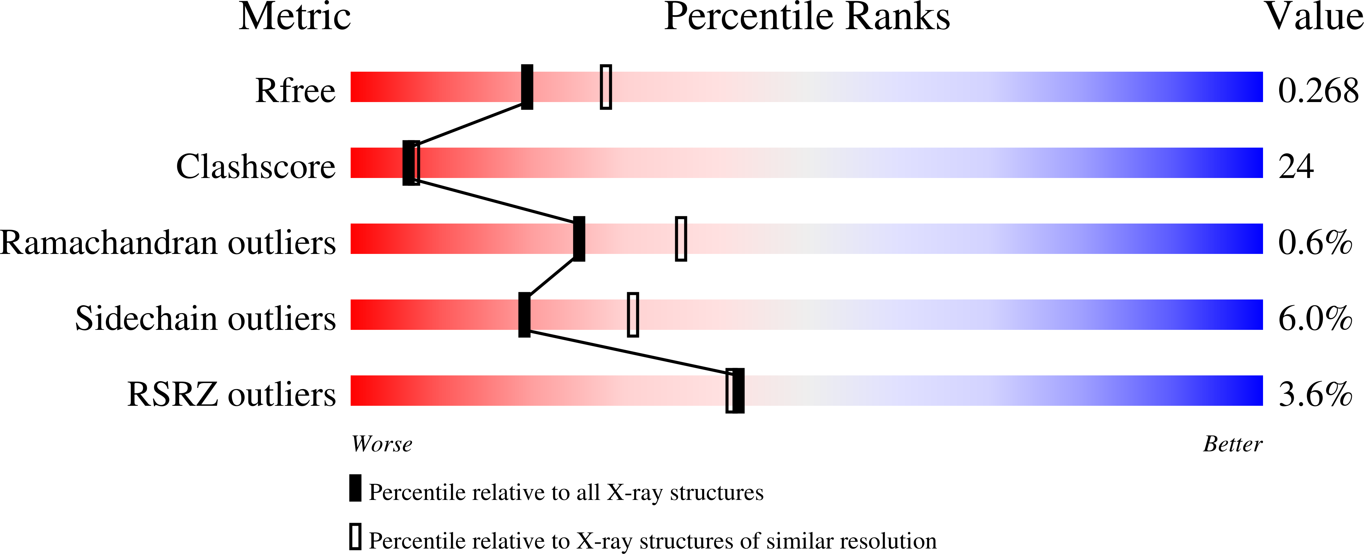
Resolution:
2.40 Å
R-Value Free:
0.26
R-Value Work:
0.24
R-Value Observed:
0.24
Space Group:
P 4 21 2
Macromolecular Entities
Structures with similar UniProt ID
Protein Blast
Polymer Type:polypeptide(L)
Molecule:Thermosome alpha subunit
Mutagens:G65C, I125T
Chain IDs:A, B, C, D
Chain Length:548
Number of Molecules:4
Biological Source:Thermococcus sp.
Ligand Molecules
Primary Citation
Crystal Structures of the Group II Chaperonin from Thermococcus strain KS-1: Steric Hindrance by the Substituted Amino Acid, and Inter-subunit Rearrangement between Two Crystal Forms.
J.Mol.Biol. 335 1265 1278 (2004)
PMID: 14729342 DOI: 10.1016/j.jmb.2003.11.028

Abstact

The crystal structures of the group II chaperonins consisting of the alpha subunit with amino acid substitutions of G65C and/or I125T from the hyperthermophilic archaeum Thermococcus strain KS-1 were determined. These mutants have been shown to be active in ATP hydrolysis but inactive in protein folding. The structures were shown to be double-ring hexadecamers in an extremely closed form, which was consistent with the crystal structure of native alpha8beta8-chaperonin from Thermoplasma acidophilum. Comparisons of the present structures with the atomic structures of the GroEL14-GroES7-(ADP)7 complex revealed that the deficiency in protein-folding activity with the G65C amino acid substitution is caused by the steric hindrance of the local conformational change in an equatorial domain. We concluded that this mutant chaperonin with G65C substitution is deprived of the smooth conformational change in the refolding-reaction cycle. We obtained a new form of crystal with a distinct space group at a lower concentration of sulfate ion in the presence of nucleotide. The crystal structure obtained at the lower concentration of sulfate ion tilts outward, and has much looser inter-subunit contacts compared with those in the presence of a higher concentration of sulfate ion. Such subunit rotation has never been characterized in group II chaperonins. The crystal structure obtained at the lower concentration of sulfate ion tilts outward, and has much looser inter-subunit contacts compared with those in the presence of a higher concentration of sulfate ion.

Legend

Protein

Chemical

Disease

Primary Citation of related structures