1Q2U image
Deposition Date 2003-07-26
Release Date 2003-10-07
Last Version Date 2024-02-21
Entry Detail
PDB ID:
1Q2U
Title:
Crystal structure of DJ-1/RS and implication on familial Parkinson's disease
Biological Source:
Source Organism:
Homo sapiens (Taxon ID: 9606)
Host Organism:
Method Details:
Experimental Method:
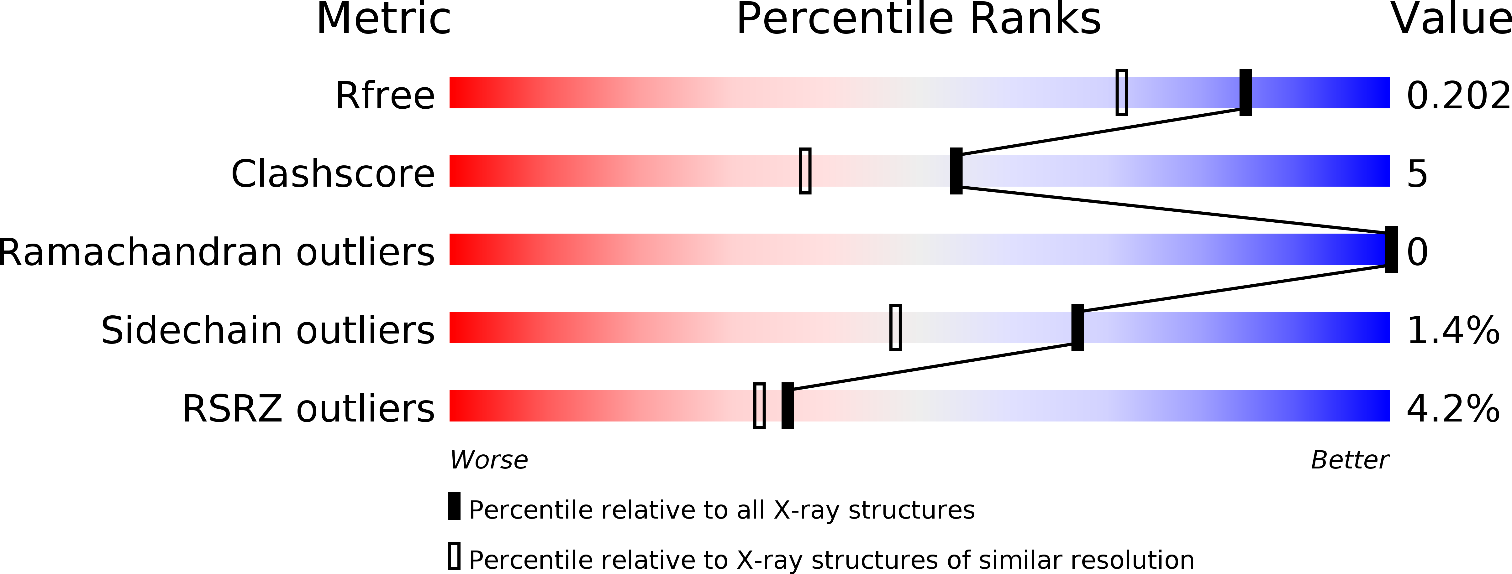
Resolution:
1.60 Å
R-Value Free:
0.20
R-Value Work:
0.19
Space Group:
P 31 2 1
Macromolecular Entities
Structures with similar UniProt ID
Protein Blast
Polymer Type:polypeptide(L)
Molecule:RNA-binding protein regulatory subunit
Gene (Uniprot):PARK7
Chain IDs:A
Chain Length:189
Number of Molecules:1
Biological Source:Homo sapiens
Primary Citation
Crystal structure of DJ-1/RS and implication on familial Parkinson's disease
Febs Lett. 549 171 175 (2003)
PMID: 12914946 DOI: 10.1016/S0014-5793(03)00764-6

Abstact

DJ-1 is a protein involved in multiple physiological processes, including cancer, Parkinson's disease, and male fertility. It is unknown how DJ-1 functions in the apparently different systems. The crystal structure of DJ-1 at 1.6 A resolution shows that DJ-1 is a helix-strand-helix sandwich and forms a dimer. The DJ-1 structure is similar to the members of the intracellular protease PfpI family. However, the catalytic triad of Cys-His-Glu is not strictly conserved in DJ-1, implying that DJ-1 has a different catalytic mechanism if it acts as a protease or DJ-1 serves as a regulatory protein in the physiological processes. The structure shows that Leu166 positions in the middle of a helix and thus predicts that the L166P mutation will bend the helix and impact the dimerization of DJ-1. As a result, the conformational changes may diminish the DJ-1 binding with its partner, leading to the familial Parkinson's disease caused by the single L166P mutation.

Legend

Protein

Chemical

Disease

Primary Citation of related structures