
Deposition Date
2003-07-18
Release Date
2003-11-18
Last Version Date
2023-08-16
Entry Detail
PDB ID:
1Q17
Keywords:
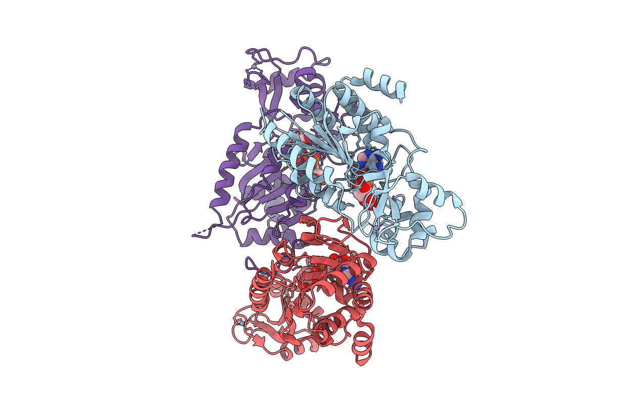
Title:
Structure of the yeast Hst2 protein deacetylase in ternary complex with 2'-O-acetyl ADP ribose and histone peptide
Biological Source:
Source Organism(s):
Saccharomyces cerevisiae (Taxon ID: 4932)
Expression System(s):
Method Details:
Experimental Method:
Resolution:
2.70 Å
R-Value Free:
0.24
R-Value Work:
0.18
R-Value Observed:
0.18
Space Group:
C 1 2 1
