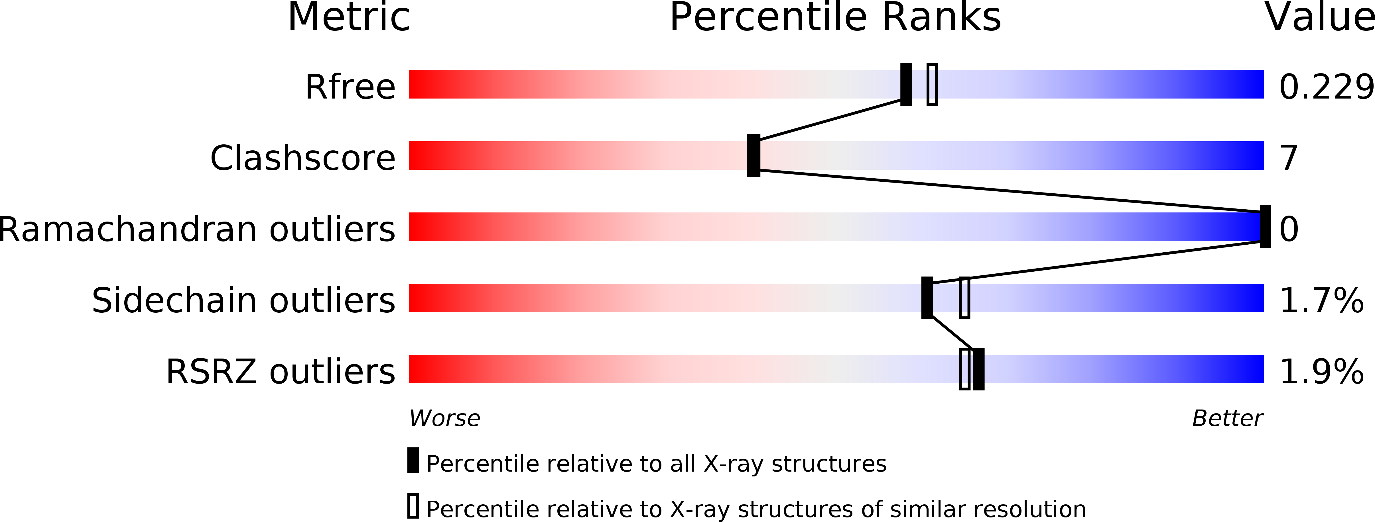
1Q0Y image
Deposition Date 2003-07-17
Release Date 2004-04-20
Last Version Date 2024-10-16
Entry Detail
PDB ID:
1Q0Y
Keywords:
Title:
Anti-Morphine Antibody 9B1 Complexed with Morphine
Biological Source:
Source Organism:
Mus musculus (Taxon ID: 10090)
Method Details:
Experimental Method:
Resolution:
2.00 Å
R-Value Free:
0.23
R-Value Work:
0.19
R-Value Observed:
0.19
Space Group:
C 1 2 1
Macromolecular Entities
Polymer Type:polypeptide(L)
Molecule:Fab 9B1, Heavy chain
Chain IDs:B (auth: H)
Chain Length:221
Number of Molecules:1
Biological Source:Mus musculus
Polymer Type:polypeptide(L)
Molecule:Fab 9B1, Light chain
Chain IDs:A (auth: L)
Chain Length:212
Number of Molecules:1
Biological Source:Mus musculus
Primary Citation
Anchoring a cationic ligand: the structure of the Fab fragment of the anti-morphine antibody 9B1 and its complex with morphine
J.Mol.Biol. 337 691 697 (2004)
PMID: 15019787 DOI: 10.1016/j.jmb.2003.12.084

Abstact

The crystal structures of an anti-morphine antibody 9B1 (to 1.6A resolution) and its complex with morphine (to 2.0 A resolution) are reported. The morphine-binding site is described as a shallow depression on the protein surface, an unusual topology for a high-affinity (Ka approximately 10(9) M(-1)) antibody against a small antigen. The polar part of the ligand is exposed to solvent, and the cationic nitrogen atom of the morphine molecule is anchored at the bottom of the binding site by a salt-bridge to a glutamate side-chain. Additional affinity is provided by a double cation-pi interaction with two tryptophan residues. Comparison of the morphine complex with the structure of the free Fab shows that a domain closure occurs upon binding of the ligand.

Legend

Protein

Chemical

Disease

Primary Citation of related structures