
Deposition Date
2003-07-10
Release Date
2004-02-24
Last Version Date
2024-10-30
Entry Detail
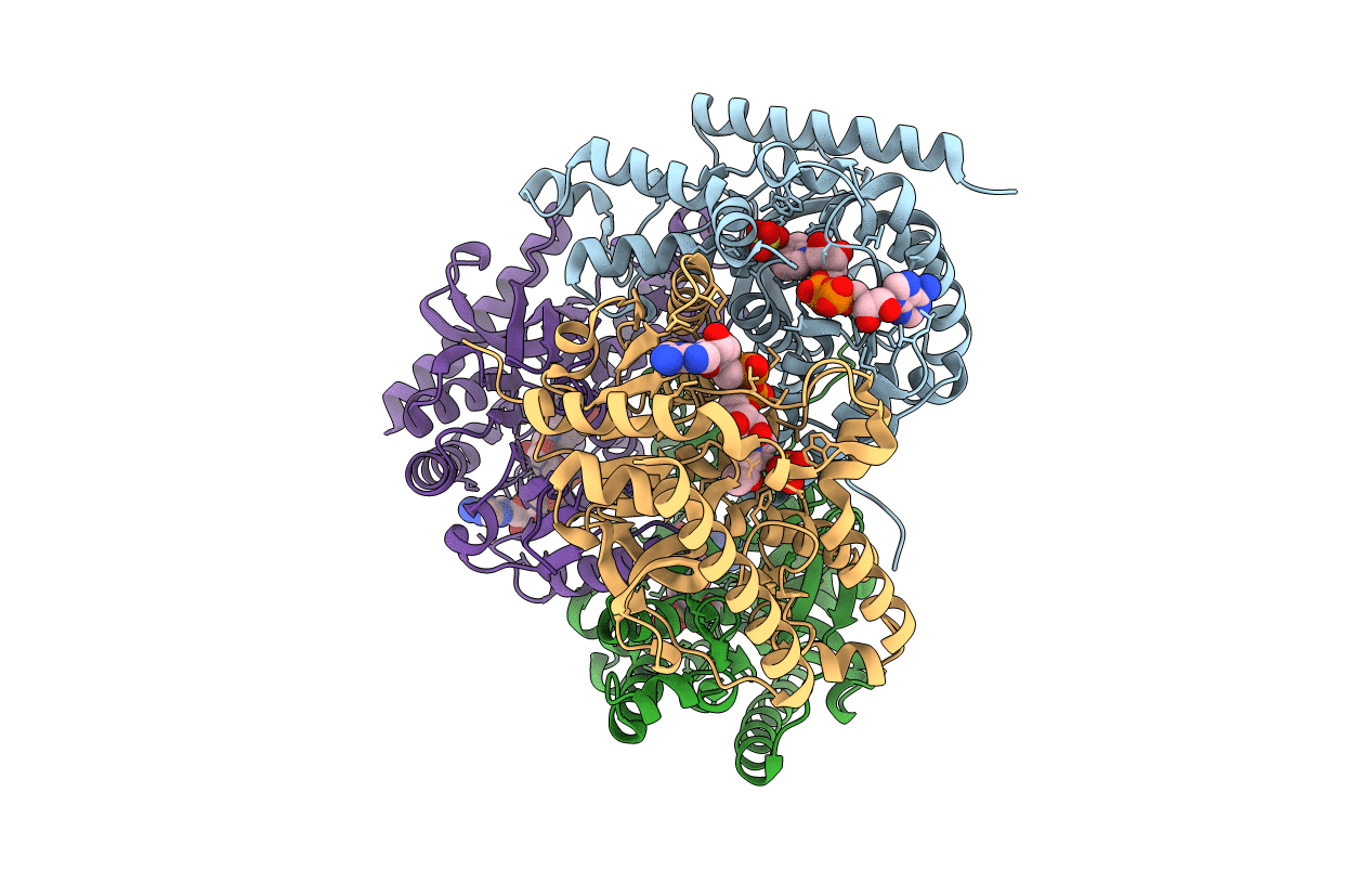
PDB ID:
1PZG
Keywords:
Title:
T.gondii LDH1 complexed with APAD and sulfate at 1.6 Angstroms
Biological Source:
Source Organism(s):
Toxoplasma gondii (Taxon ID: 508771)
Expression System(s):
Method Details:
Experimental Method:
Resolution:
1.60 Å
R-Value Free:
0.19
R-Value Work:
0.17
R-Value Observed:
0.17
Space Group:
P 1 21 1
