1PYA image
Deposition Date 1992-12-18
Release Date 1994-01-31
Last Version Date 2024-11-20
Entry Detail
PDB ID:
1PYA
Keywords:
Title:
REFINED STRUCTURE OF THE PYRUVOYL-DEPENDENT HISTIDINE DECARBOXYLASE FROM LACTOBACILLUS 30A
Biological Source:
Source Organism:
Method Details:
Experimental Method:
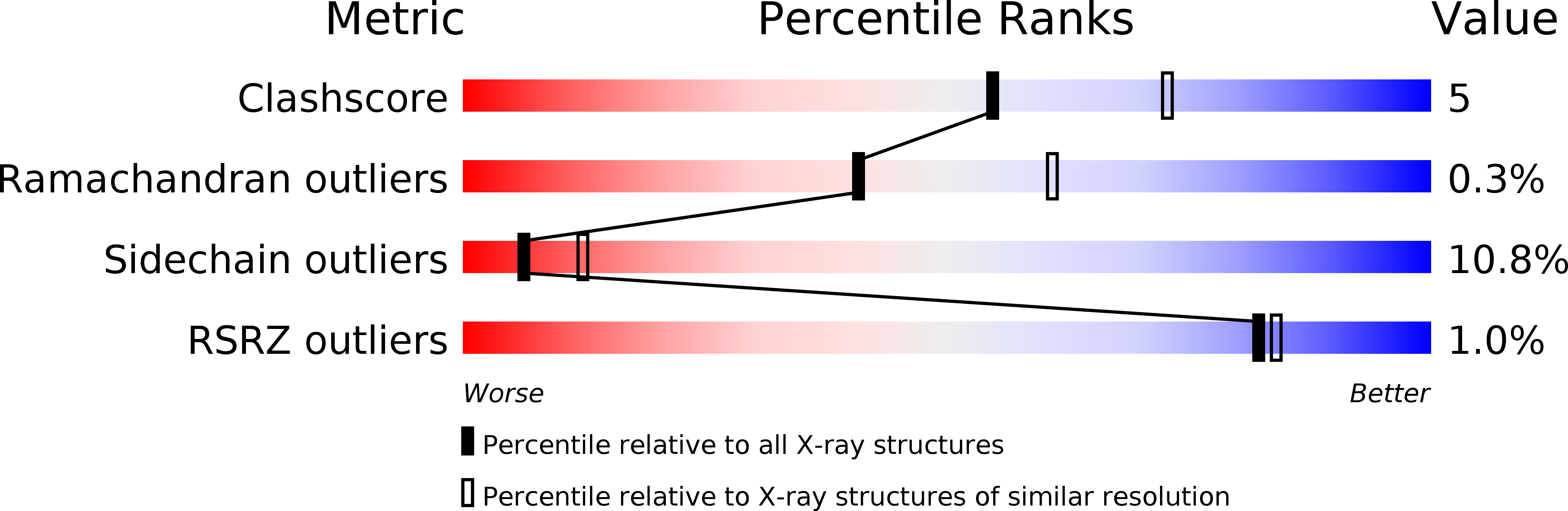
Resolution:
2.50 Å
R-Value Work:
0.15
R-Value Observed:
0.15
Space Group:
I 41 2 2
Macromolecular Entities
Structures with similar UniProt ID
Protein Blast
Polymer Type:polypeptide(L)
Molecule:PYRUVOYL-DEPENDENT HISTIDINE DECARBOXYLASE (L-HISTIDINE CARBOXYLASE)
Gene (Uniprot):hdcA
Chain IDs:A, C, E
Chain Length:81
Number of Molecules:3
Biological Source:Lactobacillus sp. 30A
Structures with similar UniProt ID
Protein Blast
Polymer Type:polypeptide(L)
Molecule:PYRUVOYL-DEPENDENT HISTIDINE DECARBOXYLASE (L-HISTIDINE CARBOXYLASE)
Gene (Uniprot):hdcA
Chain IDs:B, D, F
Chain Length:229
Number of Molecules:3
Biological Source:Lactobacillus sp. 30A
Primary Citation
Refined structure of the pyruvoyl-dependent histidine decarboxylase from Lactobacillus 30a.
J.Mol.Biol. 230 516 528 (1993)
PMID: 8464063 DOI: 10.1006/jmbi.1993.1168

Abstact

The crystal structure of the pyruvoyl-dependent histidine decarboxylase from Lactobacillus 30a has been refined to an R-value of 0.15 (for the 5.0 to 2.5 A resolution shell) and 0.17 (for the 10.0 to 2.5 A resolution shell). A description of the overall structure is presented, focusing on secondary structure and subunit association. The enzyme is a hexamer of alpha beta subunits. Separate alpha and beta-chains arise from an autocatalytic cleavage reaction between two serine residues, which results in the pyruvoyl cofactor. The central core of the alpha beta subunit is a beta-sandwich which consists of two face-to-face three-stranded antiparallel beta-sheets, flanked by alpha-helices on each side. The beta-sandwich creates a stable fold that allows conformational strain to be introduced across an internal cleavage region between the alpha and beta chains and places the pyruvoyl cofactor in a position for efficient electron withdrawal from the substrate. Three alpha beta subunits are related by a molecular three-fold symmetry axis to form a trimer whose interfaces have complementary surfaces and extensive molecular interactions. Each of the interfaces contains an active site and a solvent channel that leads from the active site to the exterior of the molecule. The trimers are related by a crystallographic two-fold symmetry axis to form the hexamer with an overall dumbbell shape. The interface between trimers has few molecular interactions.

Legend

Protein

Chemical

Disease

Primary Citation of related structures
Feedback Form
Name
Email
Institute
Feedback