Deposition Date
2003-07-08
Release Date
2003-09-30
Last Version Date
2024-10-16
Entry Detail
PDB ID:
1PY9
Keywords:
Title:
The crystal structure of an autoantigen in multiple sclerosis
Biological Source:
Source Organism(s):
Mus musculus (Taxon ID: 10090)
Expression System(s):
Method Details:
Experimental Method:
Resolution:
1.80 Å
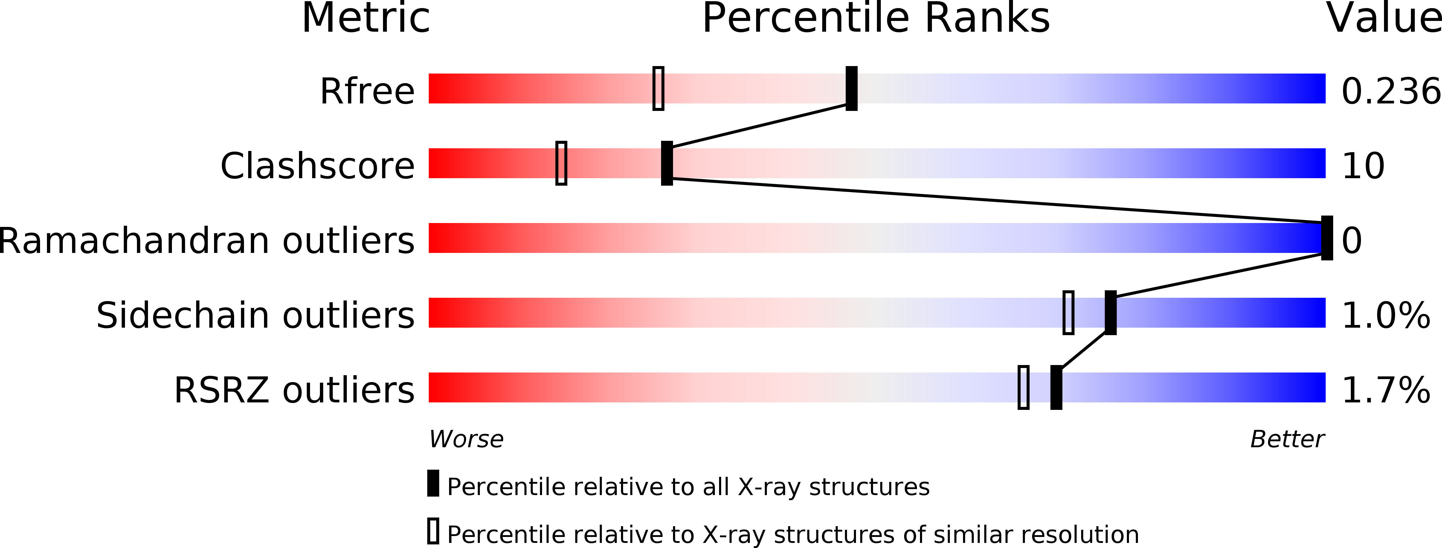
R-Value Free:
0.23
R-Value Work:
0.20
R-Value Observed:
0.20
Space Group:
I 41