1PY1 image
Entry Detail
PDB ID:
1PY1
Title:
Complex of GGA1-VHS domain and beta-secretase C-terminal phosphopeptide
Biological Source:
Source Organism:
PDB Version:
Deposition Date:
2003-07-07
Release Date:
2003-11-04
Method Details:
Experimental Method:
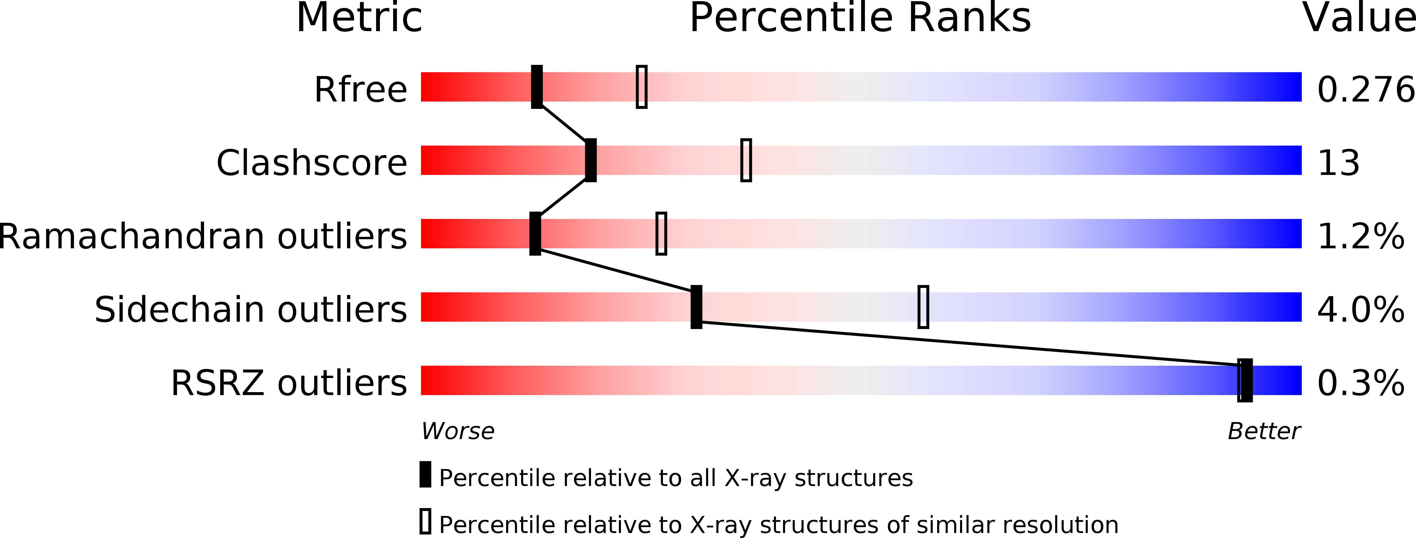
Resolution:
2.60 Å
R-Value Free:
0.28
R-Value Work:
0.23
R-Value Observed:
0.23
Space Group:
P 21 21 2
Macromolecular Entities
Polymer Type:polypeptide(L)
Description:ADP-ribosylation factor binding protein GGA1
Chain IDs:A, B, C, D
Chain Length:158
Number of Molecules:4
Biological Source:Homo sapiens
Polymer Type:polypeptide(L)
Description:Beta-secretase
Chain IDs:E, F, G, H
Chain Length:8
Number of Molecules:4
Biological Source:
Modified Residue
Compound ID Chain ID Parent Comp ID Details 2D Image
SEP E SER PHOSPHOSERINE
Primary Citation
Biochemical and structural characterization of the interaction of memapsin 2 (beta-secretase) cytosolic domain with the VHS domain of GGA proteins.
Biochemistry 42 12174 12180 (2003)
PMID: 14567678 DOI: 10.1021/bi035199h

Abstact

Memapsin 2 (beta-secretase) is a membrane-associated aspartic protease that initiates the hydrolysis of beta-amyloid precursor protein (APP) leading to the production of amyloid-beta and the onset of Alzheimer's disease (AD). Both memapsin 2 and APP are transported from the cell surface to endosomes where APP hydrolysis takes place. Thus, the intracellular transport mechanism of memapsin 2 is important for understanding the pathogenesis of AD. We have previously shown that the cytosolic domain of memapsin 2 contains an acid-cluster-dileucine (ACDL) motif that binds the VHS domain of GGA proteins (He et al. (2002) FEBS Lett. 524, 183-187). This mechanism is the presumed recognition step for the vesicular packaging of memapsin 2 for its transport to endosomes. The phosphorylation of a serine residue within the ACDL motif has been reported to regulate the recycling of memapsin 2 from early endosomes back to the cell surface. Here, we report a study on the memapsin 2/VHS domain interaction. Using isothermal titration calorimetry, the dissociation constant, K(d), values are 4.0 x 10(-4), 4.1 x 10(-4), and 3.1 x 10(-4) M for VHS domains from GGA1, GGA2, and GGA3, respectively. With the serine residue replaced by phosphoserine, the K(d) decreased about 10-, 4-, and 14-fold for the same three VHS domains. A crystal structure of the complex between memapsin 2 phosphoserine peptide and GGA1 VHS was solved at 2.6 A resolution. The side chain of the phosphoserine group does not interact with the VHS domain but forms an ionic interaction with the side chain of the C-terminal lysine of the ligand peptide. Energy calculation of the binding of native and phosphorylated peptides to VHS domains suggests that this intrapeptide ionic bond in solution may reduce the change in binding entropy and thus increase binding affinity.

Legend

Protein

Chemical

Disease

Primary Citation of related structures